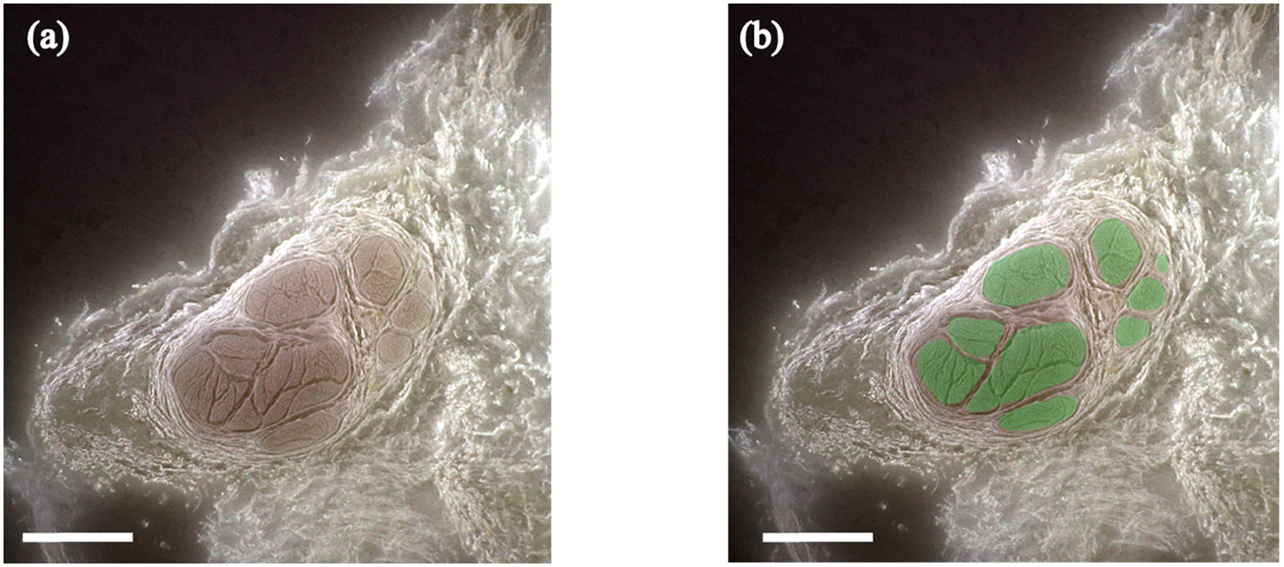
Figure 2.

3D-MUSE-cryo imaging of a fixed cadaver vagus nerve. (a) Frozen block face image, after white balancing and unsharp mask operations are applied. Fascicle structures (brown regions) can be delineated from surrounding tissue in this image. (b) Fascicle regions overlaid in green for the same block face image. Scale bar indicates 1 mm.
