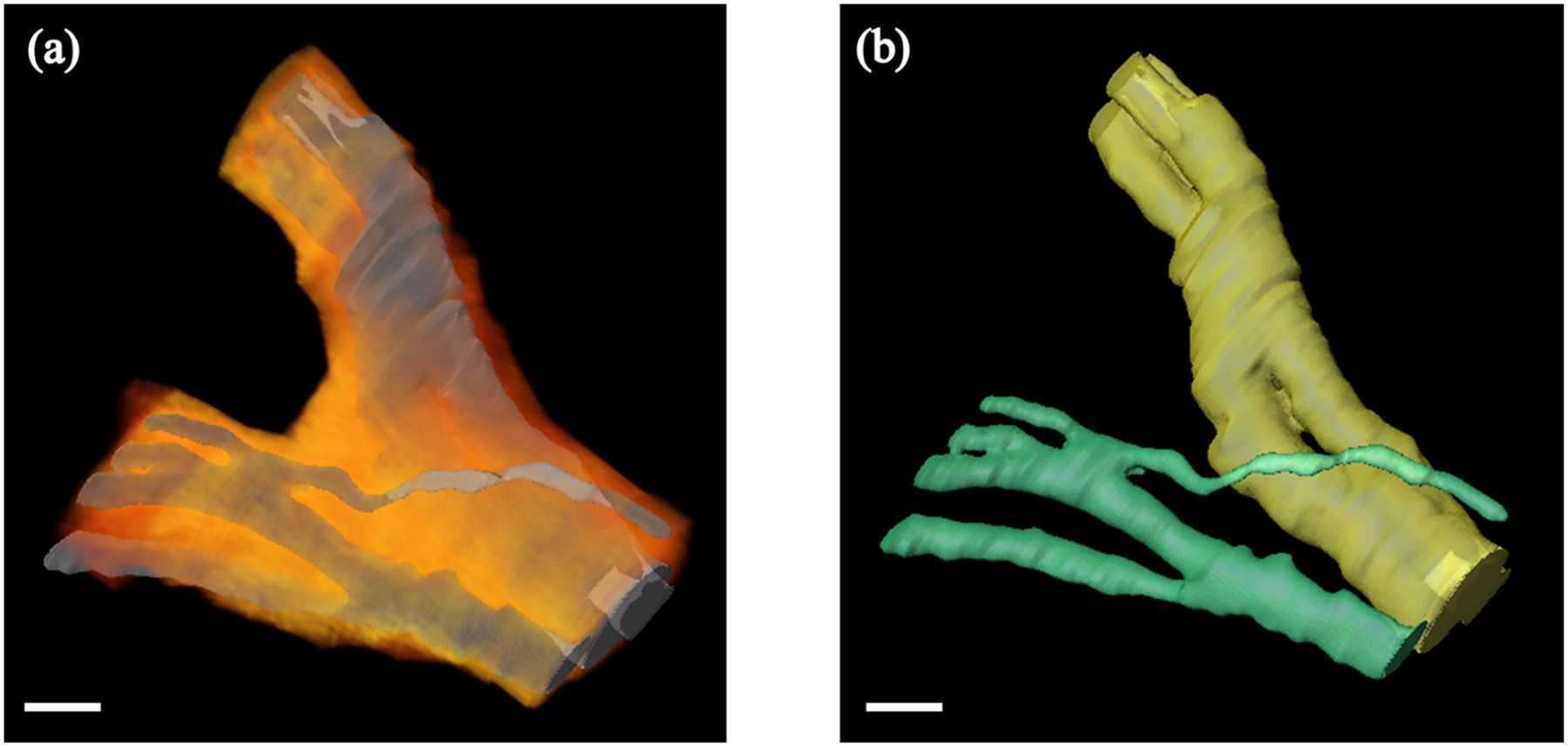
Figure 4.

3D rendering of the human vagus nerve around a branching point. (a) Fascicle labeled volumes were used to perform surface rendering (shown in gray). We visualize surrounding connective tissue (shown in an orange-red color map) by selecting pixels based on their color channel intensities and performing volume rendering. (b) Fascicles in both the main nerve trunk and the branch can be tracked individually. Fascicle surface rendering from (a) is shown, however, fascicles from the main nerve trunk and the branch are color coded in yellow and green respectively for clarity. Scale bar indicates 1 mm.
