Figure 5.
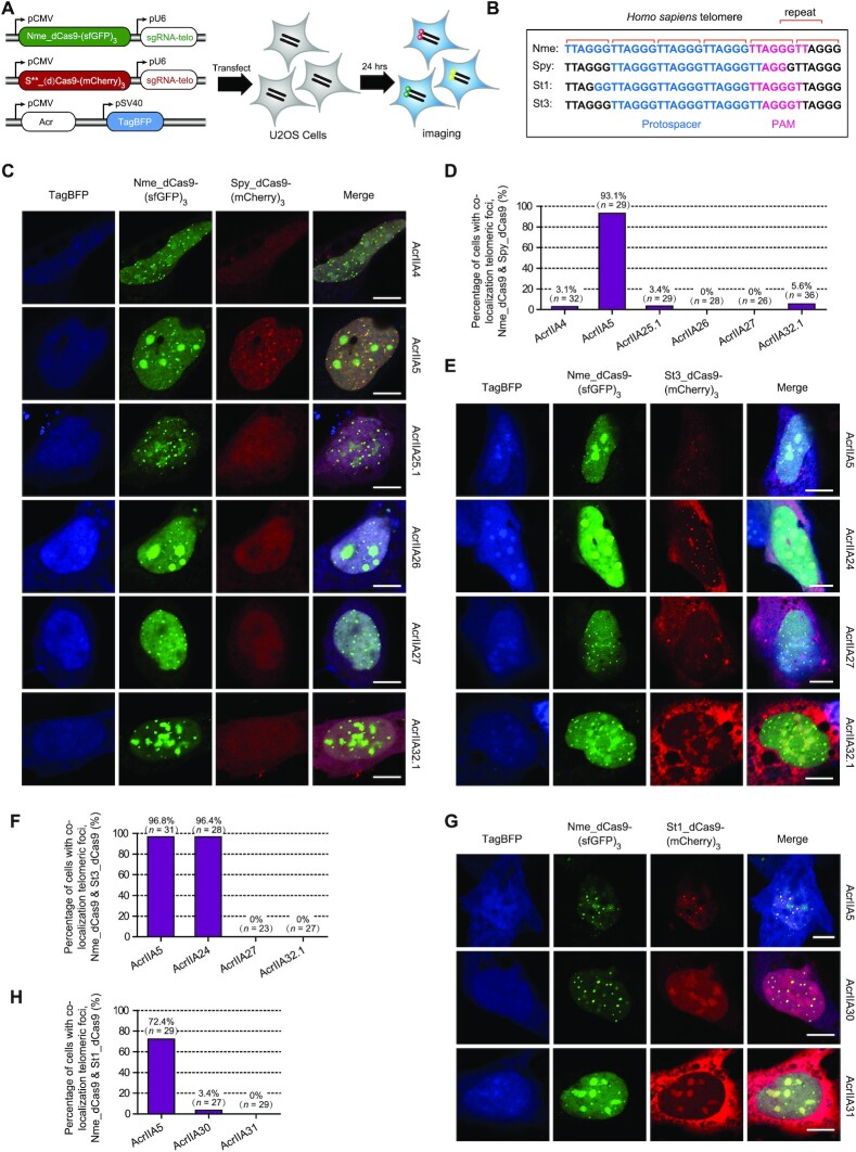
Diverse strategies employed by Acrs to disable Cas9 in human cells. (A) A schematic view of the plasmids designed for fluorescence localization of human telomeric foci to investigate the inhibitory strategy of Acrs against type II-A Cas9 orthologs. U2OS cells were co-transfected with plasmids encoding Cas9-fluorescent proteins, their respective sgRNAs targeting telomeres and Acr proteins (marked with the blue fluorescent protein TagBFP). S**_(d)Cas9-(mCherry)3 represents six plasmids used in this assay, including Spy_dCas9-(mCherry)3, Spy_Cas9-(mCherry)3, St1_dCas9-(mCherry)3, St1_Cas9-(mCherry)3, St3_dCas9-(mCherry)3 and St3_Cas9-(mCherry)3. (B) Diagrams showing targeted human telomeres in U2OS cells by Cas9 orthologs (Nme, Spy, St1 and St3) and their respective protospacer sequences. (C) Representative images of U2OS cells co-transfected with Nme_dCas9-(sfGFP)3, Spy_dCas9-(mCherry)3 and Acr plasmids. The fluorescent channels are shown at the top of the figure, and different Acr proteins are shown at the right of each row. The scale bars represent 10 μm. (D) Quantitation of Spy_dCas9-(mCherry)3 telomeric foci by calculating the percentage of cells with co-localization telomeric foci of Nme_dCas9-(sfGFP)3 and Spy_dCas9-(mCherry)3 in the presence of different Acr proteins. Foci were scored blind (see the ‘Materials and Methods’ for details). n = number of cells that were scored under each condition. Representative images of U2OS cells after transfection with St3_dCas9-(mCherry)3 (E) or St1_dCas9-(mCherry)3 (G), along with Nme_dCas9-(sfGFP)3 and different Acr plasmids. The scale bars represent 10 μm. Quantitation of St3_dCas9-(mCherry)3 (F) and St1_dCas9-(mCherry)3 (H) telomeric foci under each condition using the same method as in panel (D).

