Figure 6.
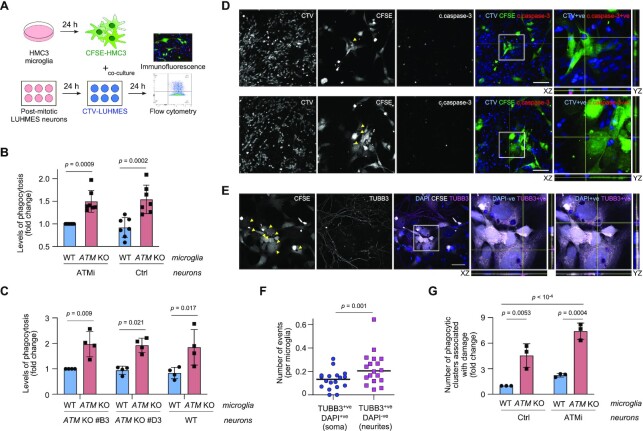
Neurotoxicity of ATM-deficient microglia is linked with aberrant phagocytosis of healthy neuronal soma and neurites. (A) Schematic of neuronal-microglial co-culture assays. Phagocytic uptake of LUHMES neurons by HMC3 microglia was analysed by flow cytometry and immunofluorescence. (B) Phagocytosis levels (CTV-stained post-mitotic LUHMES) of WT and ATM KO CFSE-labelled HMC3 cells using post-mitotic LUHMES neurons. LUHMES cells were pre-treated with AZD1390 for 3 days and released from inhibition prior to setting up the co-cultures (ATMi) or left untreated (Ctrl). Phagocytosis is relative to WT HMC3/ATMi LUHMES. Mean ± S.D. shown (n = 4). Two-way ANOVA with Sidak's multiple comparisons used. (C) Phagocytosis levels (CTV-stained post-mitotic LUHMES) of WT and ATM KO CFSE-labelled HMC3 cells using WT and two independent clones of ATMKO post-mitotic LUHMES neurons as substrates. Phagocytosis is relative to WT HMC3/ATM KO LUHMES (clone #B3), in which 9.9 ± 3.4% of cells are phagocytic. Mean ± S.D. shown (n = 4). Two-way ANOVA with Sidak's multiple comparisons used. (D, E) Representative immunofluorescence images of phagocytosis events by ATM KO microglia as in (B). Confocal Z-stack compression images and relevant XY and YZ projections shown. Scale bar: 50 μm. (D) Arrows indicate microglial phagolysosomes containing engulfed neuronal bodies (CTV+ve), which are positive (top) or negative for cleaved caspase-3 (bottom). CFSE-labelled ATM KO HMC3 microglia (CFSE; green), cleaved caspase-3 (c.caspase-3; red), CTV-labelled LUHMES neurons (CTV; blue). (E) Arrows indicate microglial phagolysosomes containing neuronal soma (TUBB3+ve, DAPI+ve) and neurites (TUBB3+ve, DAPI-ve). CFSE-labelled ATM KO HMC3 microglia (CFSE; green), β3-tubulin (TUBB3; red), DAPI (blue). (F) Quantification of phagocytic events as in (E). Distribution of TUBB3+ve DAPI+ve versus TUBB3+ve DAPI-ve events per microglia shown (n = 20 fields of view from two independent biological experiments). Wilcoxon matched-pairs signed rank test used. (G) Quantification of phagocytic microglial clusters associated with damage to the neuronal network as in (B). Data are relative to WT microglia co-cultured with untreated LUHMES post-mitotic neurons (Ctrl). Mean ± S.D. shown (n = 3). Two-way ANOVA with Tukey's multiple comparison's test used.

