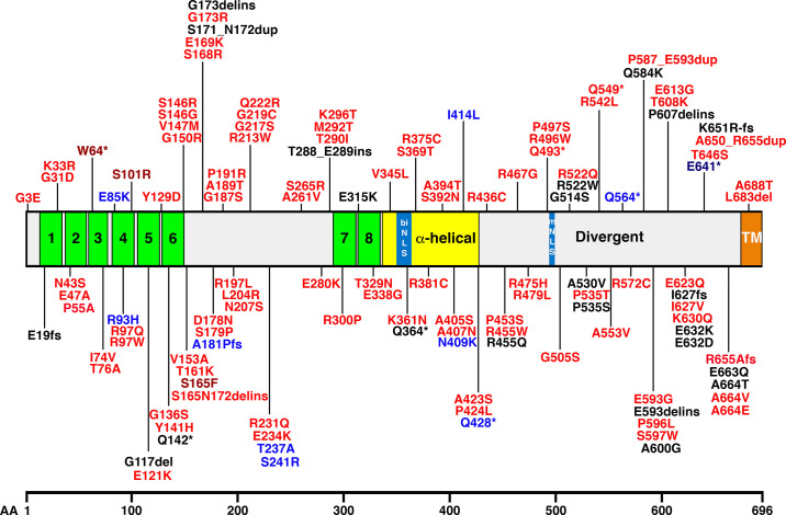
FIGURE 16.
Distribution of junctophilin 2 (JPH2) variants in ClinVar superimposed onto JPH2 protein domains. The bar in the middle of the figure represents the human JPH2 protein; functional domains are marked as follows: number green bars represent membrane occupation and recognition nexus (MORN) domains, the yellow box represents the alpha-helical domain, the divergent domain is white, and TM marks the transmembrane domain. The numbers represent amino acids residues; the letters represent amino acid identifiers. biNLS, biphasic nuclear localization signal; mNLS, monopartite nuclear localization signal; del, deletion; dup, duplication; fs, frameshift; ins, insertion. *Stop codon. Red marks hypertrophic cardiomyopathy-linked variants, and dark red indicates LP/P variants (W64*, S101R, S165F). Blue marks dilated cardiomyopathy-linked variants, dark blue indicates LP/P variant (E641*). Black marks variants with an unknown disease association.

