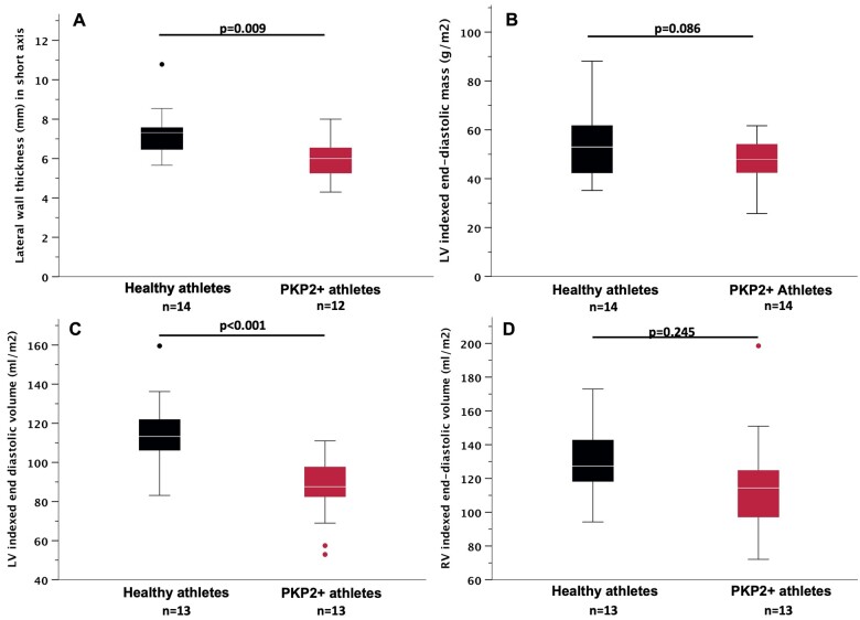
Figure 8.
Cardiac magnetic resonance in arrhythmogenic right ventricular cardiomyopathy patients and healthy controls with history of comparable athletic training. (A) Short-axis recording of lateral wall thickness. (B) Left ventricular indexed end-diastolic mass. (C) Left ventricular indexed end-diastolic volume. (D) Right ventricular indexed end-diastolic volume. Black: healthy athletes. Red: PKP2-positive athletes. Student’s t-test. Boxplots: median and interquartile range. Whiskers: 1.5× interquartile range, with outliers indicated as single dots.

