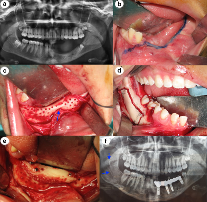
Fig. 3.
a Panoramic X-ray at the age of 16 years. b The incision line to expose the recipient side of the mandible. c Perforation of the cortical bone in the recipient site. The positions of the mental nerve are close to the residual mandibular ridge (arrow). d Intraoral photograph of the osteotomy in the donor site. e Fixation of the block graft to the ridge with three screws. f Panoramic radiograph showing the donor side (arrow) 7 months after the bone block harvest and the placement of the 4 implants

