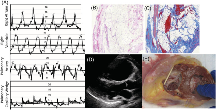
Figure 2.

Representative sample of pre‐transplant testing results and explant pathology. (A) Right heart catheterization showing elevated right‐sided filling pressures, low pulmonary artery pulse pressure, and low left atrial filling pressures. Patient had a low cardiac output and cardiac index (not shown). The vertical scale shown is in millimetres of mercury (mmHg). (B) Haematoxylin and eosin stain showing fibrofatty replacement of right ventricular myocardium at ×2 magnification. (C) Masson trichrome stain showing increased fibrosis of right ventricular myocardium at ×2 magnification. (D) Parasternal long‐axis (PLAX) echocardiography image showing dilation of right ventricle. (E) Native heart explant showing dilated right ventricle and wall thinning with fibrofatty replacement.
