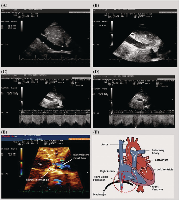
Figure 1.

Caval flow regulation natural phenomenon (CFRNP) or dynamic stenosis of the IVC. (A) Normal IVC in long‐axis view. (B) Abnormal IVC and dynamic restriction of the IVC flow. (C) Echo image showing IVC dynamic stenosis in expiration; the flow velocity is 1.8 m/s, which corresponds to 13.8 mmHg gradient. (D) Echo image showing IVC dynamic stenosis during inspiration; the flow velocity is 2.08 m/s, which corresponds to 17.3 mmHg gradient. (E) Important features of caval stenosis. (F) Schematic representation of the dynamic stenosis of the IVC.
