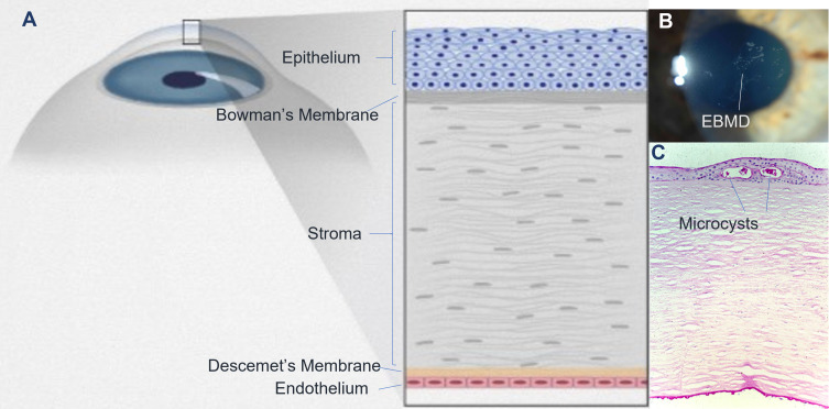
Figure 1.
Anatomy, clinical picture, and histopathology of corneal epithelial basement membrane dystrophy (EBMD). EBMD occurs at the level of corneal epithelium and basement membrane (A). Clinically, it manifests as map-dot fingerprint opacities (B). Histology shows microcystic changes of the epithelial basement membrane (C).

