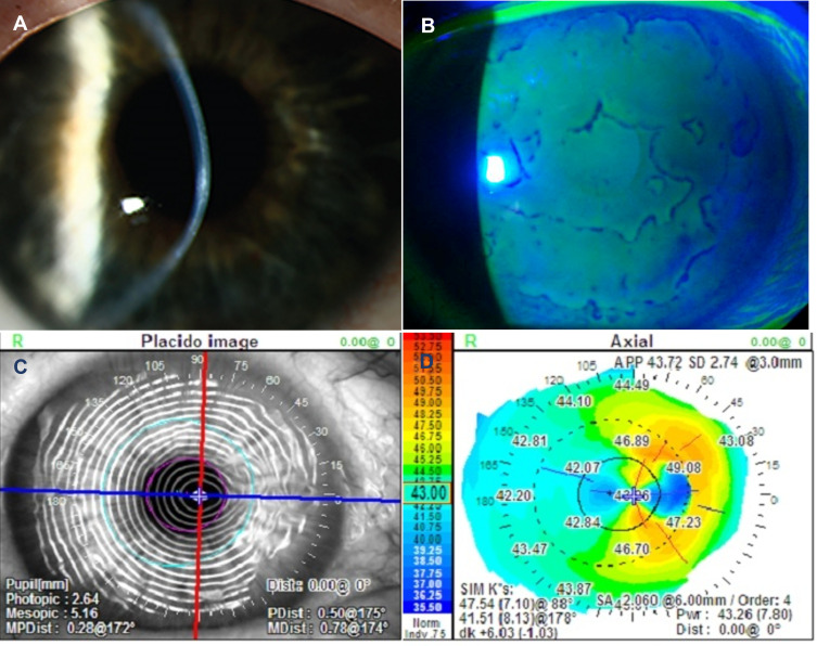
Figure 2.
Images demonstrating clinically significant epithelial basement membrane dystrophy (EBMD). Slit lamp photography showing central focal anterior corneal opacities (A) Negative fluorescein staining can identify subtle EBMD and may extend beyond the visually involved area (B). Diagnosis can be confirmed with distorted Placido rings (C) and irregular mires on corneal topography (D).

