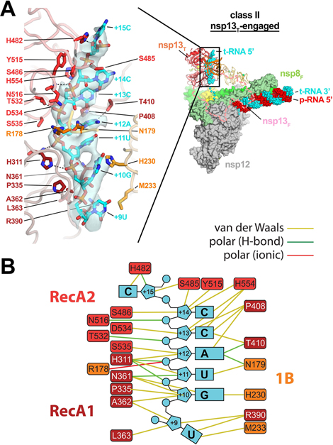
Fig. 2 |. In class II (nsp13T-engaged), the nsp13T RecA domains and 1B domain clamp onto the 5’-single-stranded t-RNA.
A. (right) Overall view of the nsp13T-engaged structure. Proteins are shown as molecular surfaces except nsp13T is shown as a backbone ribbon, and nsp13F is removed and shown only as a dashed outlline. The RNA is shown as atomic spheres. The boxed region is magnified on the left.
(left) Nsp13T is shown as a backbone worm but with side chains that interact with the t-RNA shown. Cryo-EM density for the downstream 5’-t-RNA segment is shown (transparent blue surface) with the t-RNA model superimposed. The pattern of purines/pyrimidines in the RNA density was clear and unique, allowing the identification of the sequence register for the nsp13T-bound RNA.
B. Schematic illustrating nsp13T-RNA interactions.
Also see Supplementary Video 2.

