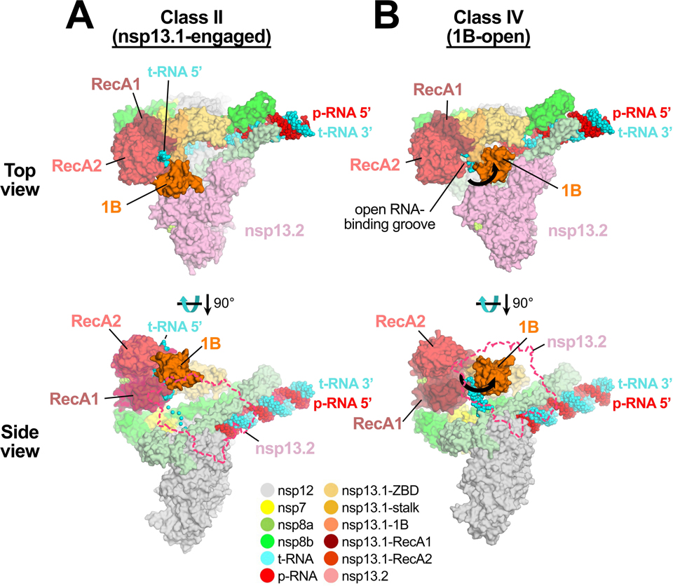
Fig. 4 |. 1B-open structure.
Comparison of nsp13T-engaged (A) and 1B-open (B) structures. Two views are shown, a top view (top) and a side view (bottom). In the top view, the proteins are shown as molecular surfaces and color-coded according to the key at the bottom. In the side view, nsp13F is shown only as a dashed outline. The RNA is shown as atomic spheres. In the 1B-open structure (B), the nsp13T 1B domain is rotated open by 85° (represented by thick black arrows). The 5’-t-RNA emerging from the RdRp active site approaches the nsp13T RNA binding groove but does not enter it.
Also see Supplementary Video 2.

