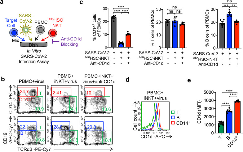
Fig. 3.
AlloHSC-iNKT cells reduce virus-infection promoted inflammatory monocytes. a Experimental design. 293T-ACE2-FG cells were infected by SARS-CoV-2 virus. After 1 day, ATO culture-generated AlloHSC-iNKT cells and donor-mismatched PBMCs were added and incubated for 24 h. Flow cytometry was used to detect cell populations. b FACS detection of CD14+ monocytes, T cells, and B cells in PBMCs. c Quantification of b (n = 5). d FACS detection of CD1d expression on CD14+ monocytes, T cells, and B cells. e Quantification of d (n = 5). Representative of 3 experiments. Data are presented as the mean ± SEM. ns, not significant, *P < 0.05, **P < 0.01, ***P < 0.001, ****P < 0.0001, by 1-way ANOVA

