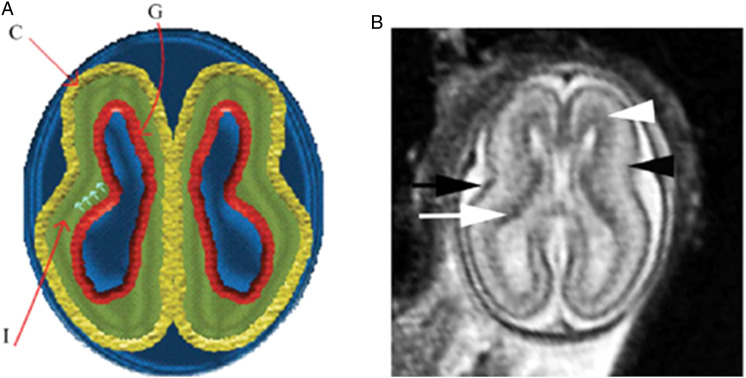
Figure 2.
Normal multi-layered magnetic resonance imaging (MRI) appearance of fetal brain early in gestation. (A) A diagram representing the fetal brain at 19 weeks of gestation shows surface and multi-layered appearance of the parenchyma with an inner germinal matrix (G), intermediate layer (l), and a developing cortex. (C). The small arrows point to the direction of the migrating neurons from germinal matrix to the developing cortex. (B) Axial balanced fast field echo MR image of a normal brain at 19 weeks of gestation shows a smooth surface and multi-layered parenchyma with an inner hypointense germinal matrix (white arrow), an intermediate layer, and an outer hypointense developing cortex (black arrow). Two additional sublayers can be identified: subventricular zone (white arrowhead) and subplate (black arrowhead). Subventricular zone is thick in the frontal region and shows slightly hypointense signal as it contains germinal matrix with increased cell pro-duction. The subplate zone appears slightly hyperintense as it has high water content, because of extracellular matrix. Reproduced by permission from SAGE Publications (Saleem 2013).

