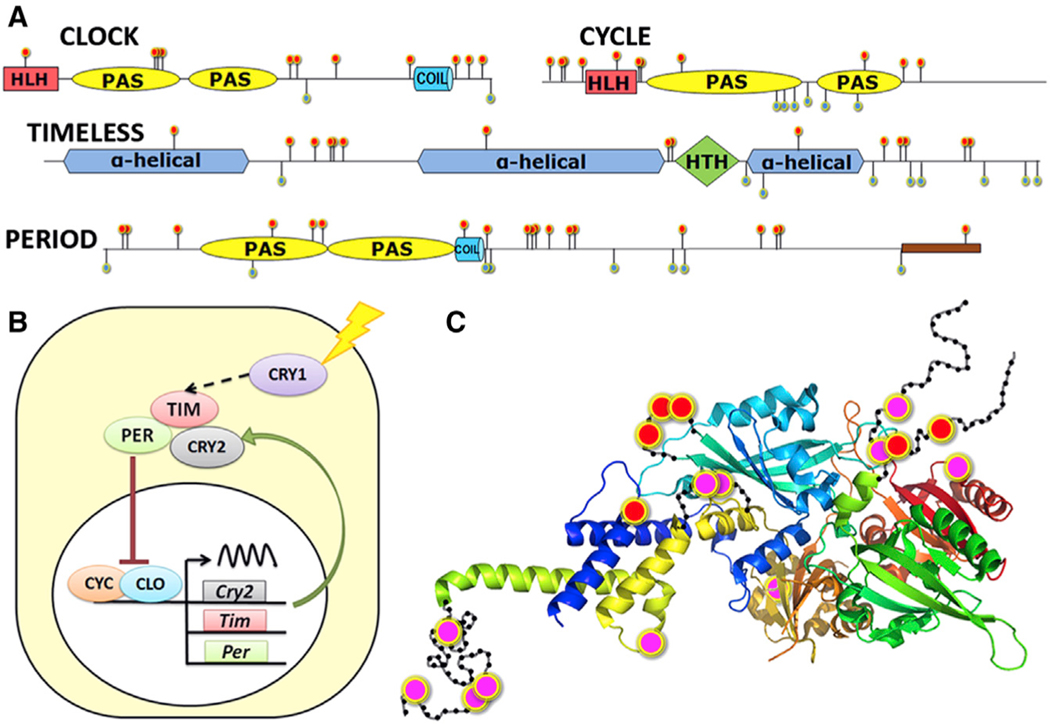
Figure 4. Circadian Clock System Could Explain Differences in Diapause between Pgl and Pca.
(A) Domain diagram of CLOCK, CYCLE, PERIOD, and TIMELESS. Mutations within species are marked by green flags, and positions that are conserved within but differ between species are marked by red flags.
(B) Circadian clock system. CRY, cryptochrome proteins.
(C) Map of inter-species mutations on the spatial structure template (PDB id: 4F3L) of CLOCK/CYCLE complex. The mutations are marked by red (CLOCK) and pink (CYCLE) dots, and the approximate position of disordered loops is shown as black beads on threads.
See also Figure S5.

