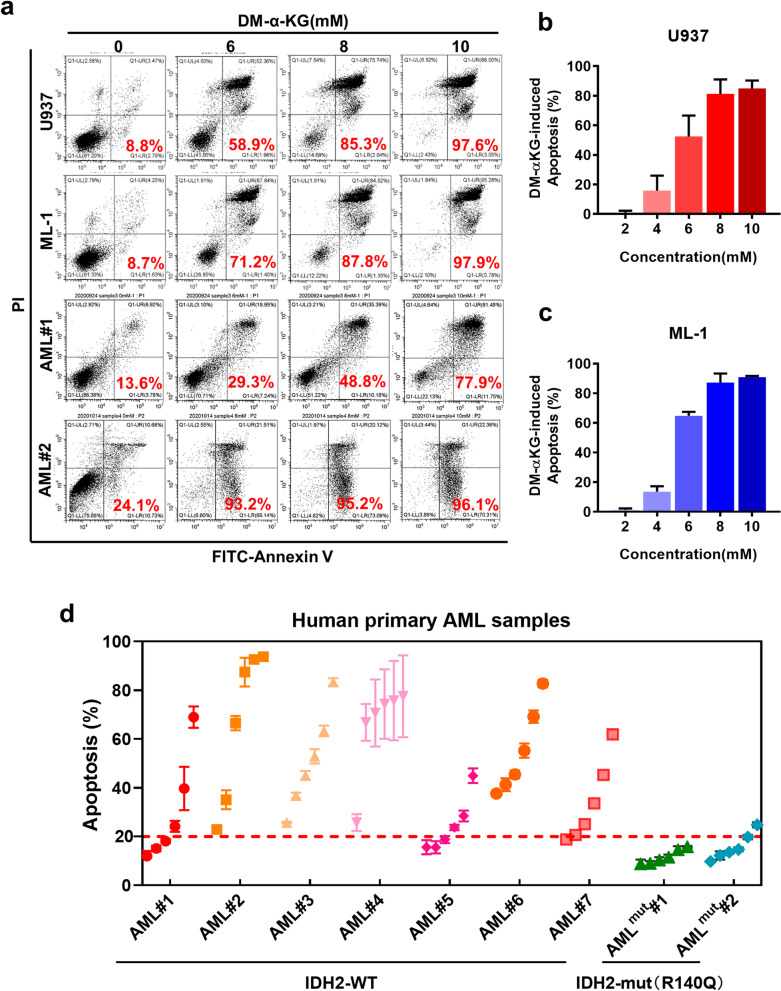
Fig. 5.
Cytotoxic effect of α-KG in AML cells with wild-type IDH2. a Induction of apoptosis by cell-permeable DM-αKG in AML cell lines (U937 and ML-1) and primary AML cells isolated from patients with wt-IDH2 (AML#1 and AML#2). Cells were treated with the indicated concentrations of DM-αKG for 48 h, and apoptosis was measured by flow cytometry analysis of annexin-V positivity. The number inside each panel shows the percentage of dead cells. b, c Quantitation of the concentration-dependent apoptosis induced by DM-αKG in U937 and ML-1 cells (n = 3, mean ± SD). d Apoptosis of human primary AML cells harboring wild-type (n = 7) or mutant IDH2 (IDH2-R140Q, n = 2) treated with various concentrations (2, 4, 6, 8 and 10 mM) of DM-αKG for 48 h. Apoptosis was measured by flow cytometry analysis of annexin-V positivity

