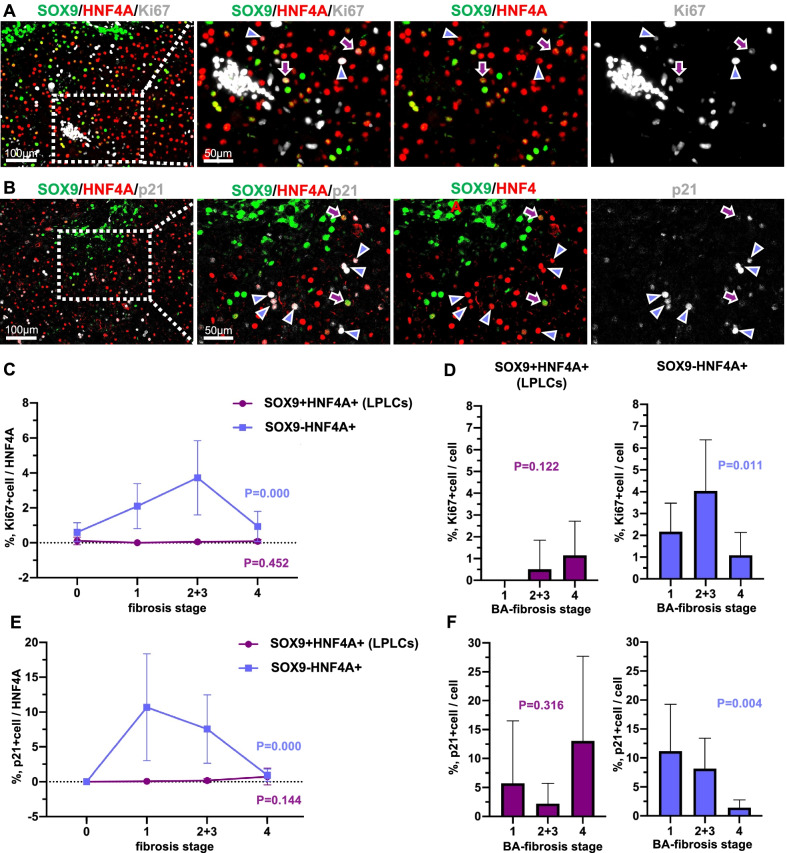
Fig. 3.
LPLCs exhibit superior proliferation and anti-senescence abilities as compared to SOX9-negative hepatocytes with the progression of cholestasis. Multiple immunofluorescence staining of LPLCs with (A) proliferation marker (Ki67) and (B) anti-senescence marker (p21). Overall percentage (C) and cell cluster percentage (D) of Ki67+ LPLCs and Ki67+ SOX9-negative hepatocytes in different fibrosis stages. Overall percentage (E) and cell cluster percentage (F) of p21+ LPLCs and p21+ SOX9-negative hepatocytes in different fibrosis stages

