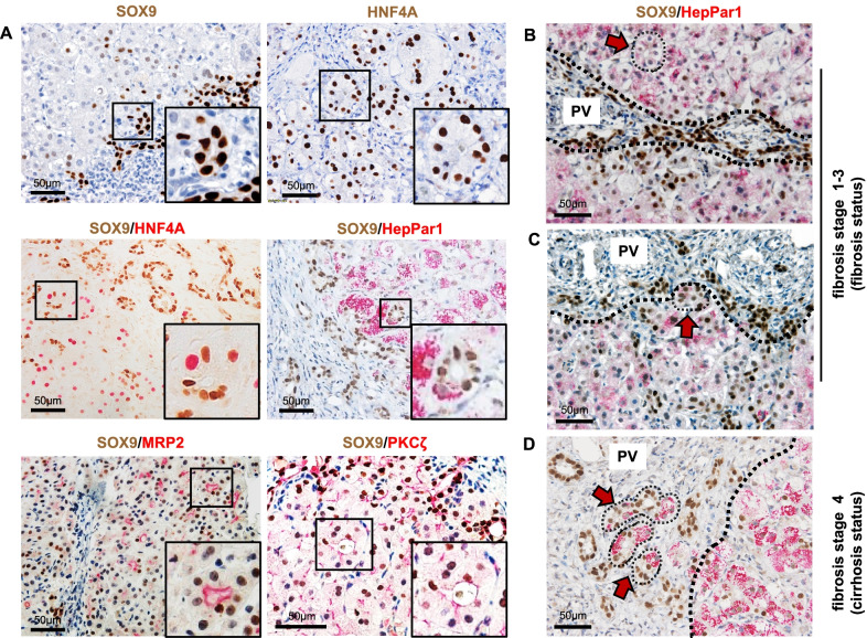
Fig. 4.
LPLCs differentiated into RDCs in the periportal region with the progression of cholestasis. A Pseudo-rosette formation in the periportal parenchyma appears to have bipotent characteristics during cholestatic liver damage, which co-stains with the LPLC markers SOX9 and HNF4A and with other hepatocyte and cholangiocyte markers, namely HepPar1, MRP2, and PKCζ. Pseudo-rosette hepatocytes (HepPar1-positive) appeared (B) in the periportal parenchyma with weak SOX9 staining and (C) in the adjacent portal region with increasing SOX9 staining and ductular-like structure in the fibrosis stage and (D) occurred inside the portal region with SOX9-positive ductular structure in the cirrhosis stage

