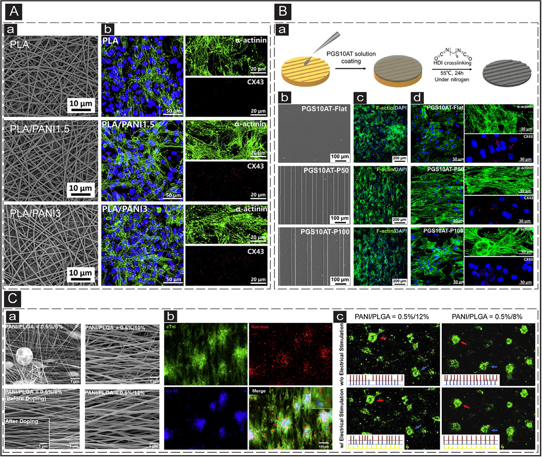
Fig. 5.

ECPs application in cTE. (A) Electrospun nanofibrous constructs for cTE. a: SEM images of the electrospun PLA scaffolds with different PANI contents. b: IF images show the cardiac-specific markers (Cx-43 (red) and sarcomeric α-actinin (green)) expression in the CMs cultured on the electrospun scaffolds with various PANI contents. Adapted with permission from [171]. Copyright © 2017, Elsevier. (B) Micropatterned electroconductive PGS-aniline scaffolds for cTE. a: Schematic illustrating the process of PGS10AT-6H micropatterns. b: SEM images of the patterned PGS10AT films. c: IF images show F-actin (green) expression in the CMs cultured on patterned scaffolds after two days. d: IF images of the expression of cardiac-specific markers (α-actinin (green) and Cx-43 (red)) in the CMs cultured on the scaffolds after eight days. Adapted with permission from [190]. Copyright © 2019, Elsevier. (C) Electrical coupling of separated CMs cultured on electrospun conductive PANI/PLGA nanofibrous scaffolds. a: SEM images of the electrospun PANI/PLGA scaffolds with various ratios of PANI/PLGA (w/v). b: IF images of the expression of cardiac-specific markers (cTnI (green) and Cx-43 (blue)) in the CMs cultured on the doped fibrous scaffolds. c: Beating frequencies of the CMs clusters cultured on the low (PNAI/PLGA = 0.5/12%) and high (PNAI/PLGA = 0.5/8%) conductive scaffolds prior and after electrical stimulation. The red, blue, and yellow signals correspond to the red arrow, blue arrow, and stimulated electrical signals. Adapted with permission from [189]. Copyright © 2013, Elsevier.
