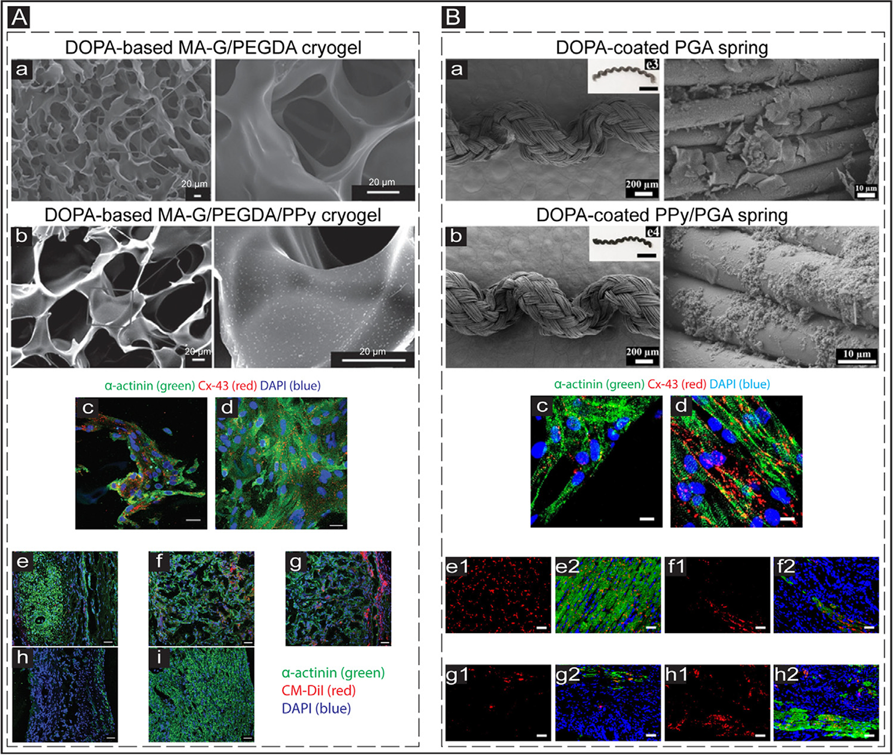
Fig. 6.

In vivo application of conductive biomaterials for cardiac regeneration after MI. (A) a&b: Scanning electron microscopy (SEM) images showing the structure of conductive and non-conductive cryogels. The PPy concentration in the conductive cyogel (b) was 2 mg/mL. The scale bar is 20 μm. c&d: IF images exhibit the expressed cardiac-specific markers (α-actinin (green) and Cx-43 (red)) in the CMs cultured on DOPA-based MA-G/PEGDA (c) and DOPA-based MA-G/PEGDA/PPy (d) at day 8 in vitro. The scale bar is 50 μm. e-i: IF images showing the expression of CM-DiI labeled (red) CMs and α-actinin (green) in the infarcted hearts treated with DOPA-based MA-G/PEGDA/PPy patch. e: Infarcted myocardium; f: Middle part of the transplanted conductive patch; g: External region of the transplanted patch; h: Infarct zone of the myocardium of the treated animals; i: Healthy tissue of the myocardium (control). The scale bar is 50 μm. Adapted with permission from [221]. Copyright © 2016, John Wiley and Sons. (B) a&b: SEM images showing the structure of non-conductive (DOPA-coated PGA) and conductive (DOPA-coated PPy/PGA) biosprings. c&d: Expressed cardiac-specific markers (α-actinin (green) and Cx-43 (red)) in the CMs cultured on DOPA-coated PGA (c) and DOPA-coated PPy/PGA (d) springs at day 7 in vitro. The scale bar is 10 μm. e-h: Images showing the cardiac-specific markers (α-actinin (green) and Cx-43 (red)) expression in the hearts of treated animals. e1&e2: Sham group; f1&f2: Infarcted group; g1&g2: Non-conductive spring (DOPA-coated PGA) group; h1&h2: Conductive spring (DOPA-coated PPy/PGA) group. The scale bar is 50 μm. Adapted with permission from [224]. Copyright © 2019, American Chemical Society.
