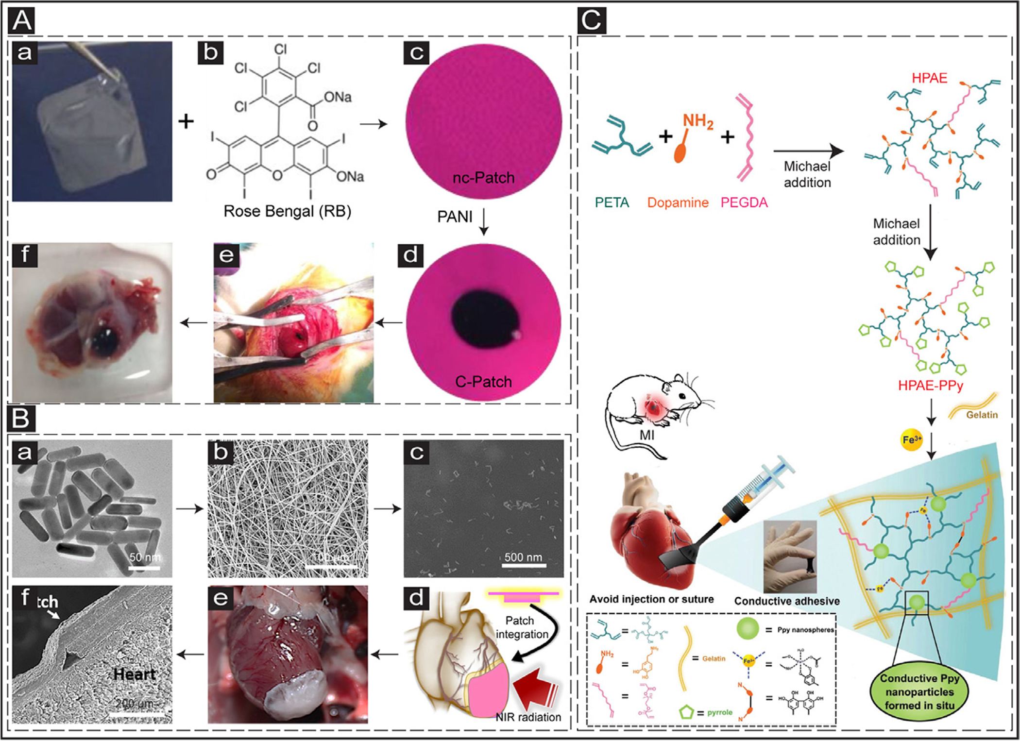
Fig. 7.

Suture-free methods for the delivery of cardiac patches in vivo. (A) a: CHI film. b&c: Rose Bengal is added to the CHI film making a non-conductive photoactivated patch (nc-Patch). d: PANI is immobilized into the nc-Patch, making a conductive patch (c-Patch). e: Transplantation of the c-Patch into the rat heart through photoadhesion. f: Explanted heart after two weeks in vivo. Adapted with permission from [212]. Copyright © 2016, American Association for the Advancement of Science. (B) Suture-free GNR-based cardiac patch. a: Synthesized GNRs. b: SEM image of electrospun albumin scaffolds. c: SEM image of incorporated GNRs into the fibrous scaffolds. d: Scheme illustrating the transplantation of cardiac patch into the beating heart by NIR radiation. e: Integration of the cardiac patch with the rat heart. f: SEM image of the integrated patch and the heart tissue. Adapted with permission from [235]. Copyright © 2018, American Chemical Society. (C) Schematic showing the development of a paintable conductive cardiac patch. Adapted with permission from [234]. Copyright © 2018, John Wiley and Sons.
