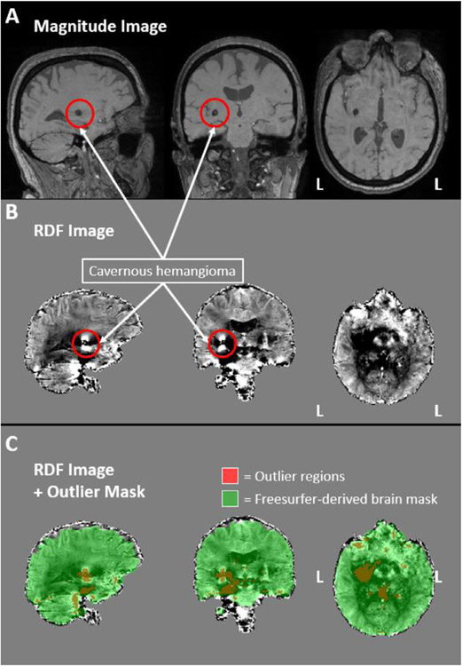
Fig. 4.
Ironsmith-based phase image outlier detection. The magnitude (panel A) and RDF (panel B) images of a participant with a clinically-confirmed cavernous hemangioma are presented, outlined with red circles. Panel C. depicts, in red, the regions identified by Ironsmith as outliers using the MAD-based outlier detection process. The green mask in panel C represents a Freesurfer-derived brain mask used to constrain the outlier detection process.

