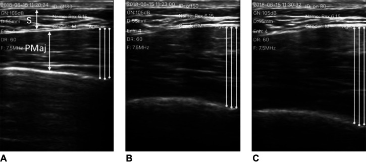
Figure 4.

An example of ultrasound images illustrating the PMaj in transverse view in 1 subject (S: subcutaneous soft tissue; PMaj: pectoralis major): (A) The PMaj at rest. (B) Maximally contracted PMaj without RUSI biofeedback. (C) Maximally contracted PMaj with RUSI biofeedback. RUSI = real-time ultrasound imaging.
