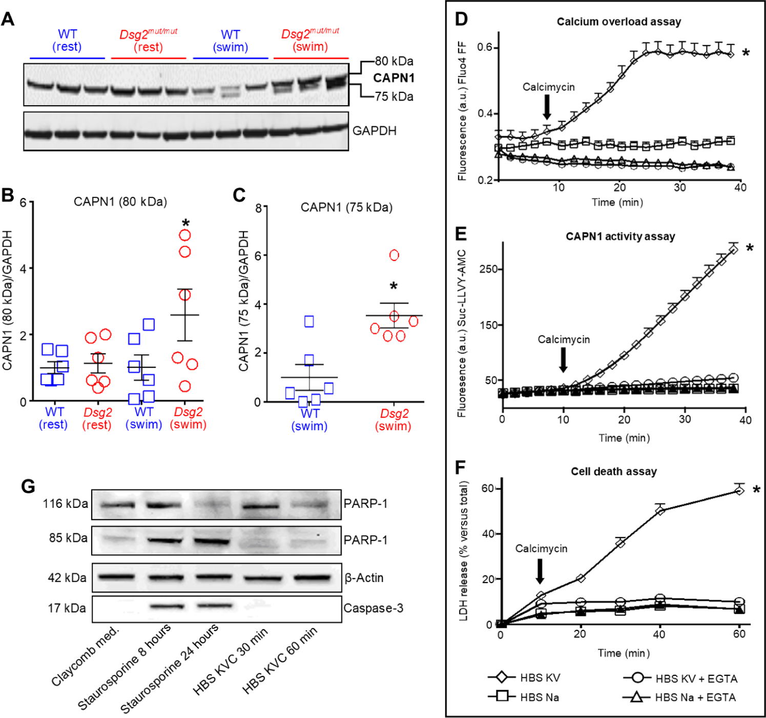
Fig. 2. CAPN1 activation is associated with calcium overload and cell death, promoting myocyte necrosis.

(A) Representative calpain-1 (CAPN1 recognizing domain IV) immunoblot of heart lysates from sedentary (rest) and exercised (swim) mice. (B and C) Quantification of CAPN1 in hearts from Dsg2mut/mut and WT mice. Data are presented as means ± SEM [n = 6 mice per genotype per cohort with *P < 0.05 Dsg2mut/mut (swim) compared to WT (swim) using one-way ANOVA in (B) and *P < 0.05 Dsg2mut/mut (swim) compared to WT (swim) using two-tailed, paired t test in (C)]. (D) HL-1 cells incubated in HBS sodium (HBS Na) medium or HBS with potassium and vanadate (HBS KV) medium in the absence or presence of 5 mM EGTA and calcimycin (1 μM, black arrow) to induce calcium (Ca2+) overload. a.u., arbitrary units. (E) CAPN1 activity, monitored by proteolytic cleavage of a synthetic peptide (Suc-LLVY-AMC) to produce fluorescence, in HL-1 cells exposed to the indicated media. (F) Death of HL-1 cells exposed to the indicated media, detected by lactose dehydrogenase (LDH) release into the media. (G) Representative immunoblot of poly (ADP-ribose) polymerase-1 (PARP-1) and caspase-3 and their cleaved product in HL-1 lysates. Cells were exposed to calcimycin in HBS KV medium (HBS KVC) or to staurosporin in the Claycomb medium (Sigma-Aldrich, no. 51800C) for the times indicated in the panel (8 or 24 hours for staurosporine; 30 or 60 min for HBS KVC). Data are representative of one of six experiments. For (D) to (F), data are presented as means ± SD (n = 6 independent experiments per cohort, with n = 3 cell culture replicates per condition; *P < 0.05 HBS KV compared to all other conditions using one-way ANOVA).
