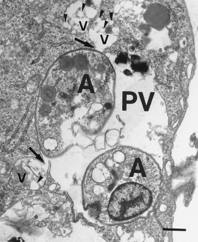
FIG. 2.
Electron micrograph showing the fusion of PLA nanoparticles with the parasitophorous vacuole. Infected macrophages were cultivated for 6 h in the presence of PLA nanoparticles and then processed for electron microscopy. PLA nanosphere-containing vacuoles (v) and one parasitophorous vacuole (PV) containing two amastigotes (A) are shown. Arrows indicate the fusion of PLA-containing vacuoles with the parasitophorous vacuole membrane. Arrowheads point to individual nanospheres. Bar, 0.5 μm.

