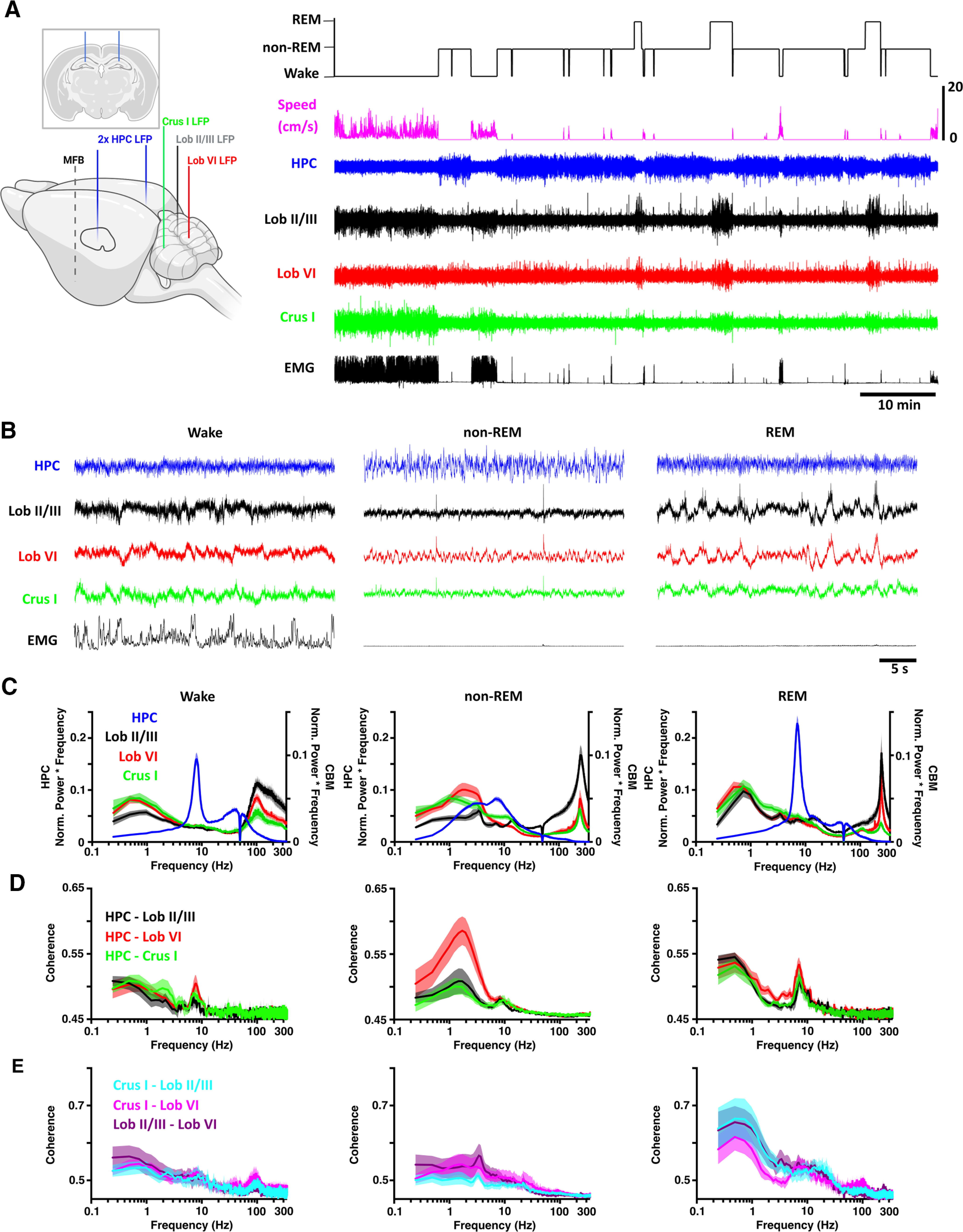
Figure 1.

Sleep state-specific activity patterns are present in the cerebello-hippocampal network. A, Left, Schematic illustration of all electrode implantation locations with coronal schematic of hippocampal electrodes as inset. Right, Hippocampal (HPC) and cerebellar cortical (Crus I, Lob VI, and Lob II/III) LFPs were recorded as mice cycled between defined wake, non-REM, and REM states. Created with www.BioRender.com. B, During wake, hippocampal theta and cerebellar <1 Hz and high γ (100-160 Hz) oscillations occurred concomitantly. Similarly, during REM, hippocampal theta oscillations were accompanied by widespread δ (<4 Hz) and very fast (∼250 Hz) cerebellar oscillations. During non-REM, high-amplitude hippocampal activity co-occurred with both slow, phasic, and very high-frequency (∼250 Hz) cerebellar oscillations. C, Mean power spectra for each of the defined states (HPC, n = 20; Crus I, n = 11; Lob VI, n = 15; Lob II/III, n = 11). D, Mean coherence between HPC-Crus I (n = 11), HPC-Lob VI (n = 15), and HPC-Lob II/III (n = 11) across states. E, Intracerebellar δ coherence is highest during REM sleep. δ band (<4 Hz) coherence was elevated during REM sleep compared with both awake and non-REM in all combinations of cerebellar recordings (Crus I-Lob II/III, n = 8; Crus I-Lob VI, n = 7; Lob II/III-Lob VI, n = 7). Shading represents SEM calculated across animals.
