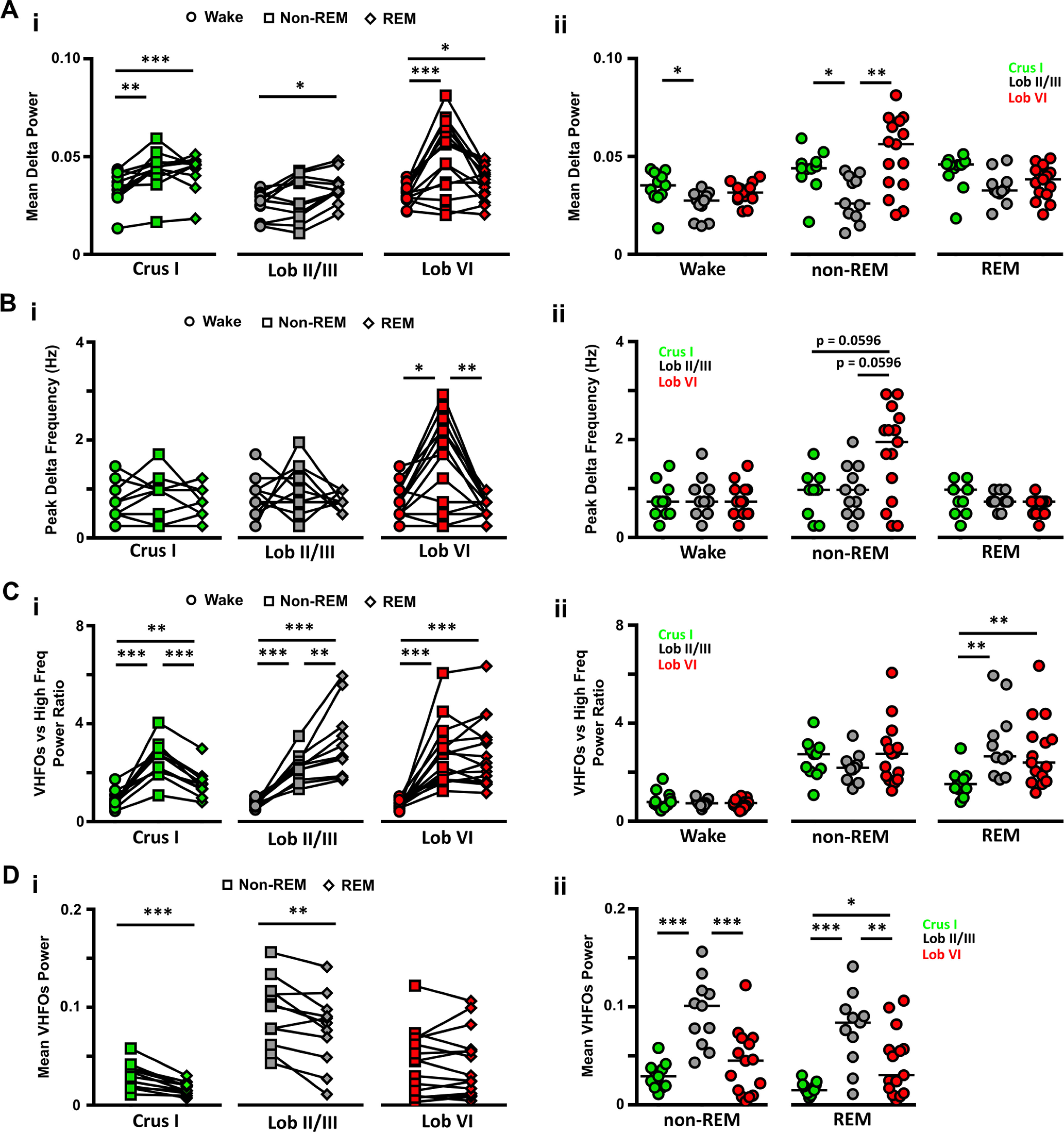
Figure 2.

LFP power in δ (0.1-4 Hz) and VHFO (240-280 Hz) frequency bands during wake and sleep. Ai, The power of δ oscillations varied between wake and the different sleep states within the three cerebellar regions. Aii, Similarly, differences between cerebellar regions were observed both during wake. Bi, The peak δ band frequency differed across sleep states only in Lob VI. Bii, Differences in the peak δ frequency band across cerebellar regions were also restricted to non-REM epochs. Ci, During wake, the power of VHFOs was lower compared with high-frequency oscillations in all regions, as indicated by the mean values <1. In contrast, during sleep, across all recorded cerebellar regions, the ratio of VFHO to high-frequency oscillation power increased significantly compared with wake (shifted to values >1), and also differed between sleep states. Cii, Significant differences across cerebellar regions were restricted to REM epochs when Crus I showed significantly smaller ratios than the other two regions. Di, VHFO power was significantly reduced during REM sleep compared with non-REM in Crus I and Lob II/III but remained unchanged in Lob VI. Dii, VHFO power was significantly higher in Lob II during both non-REM and REM sleep. For Crus 1, n = 11; Lob VI, n = 15; Lob II/III, n = 11. For detailed statistical comparisons, see Extended Data Figure 2-1. *p < 0.05. **p < 0.01. ***p < 0.001.
