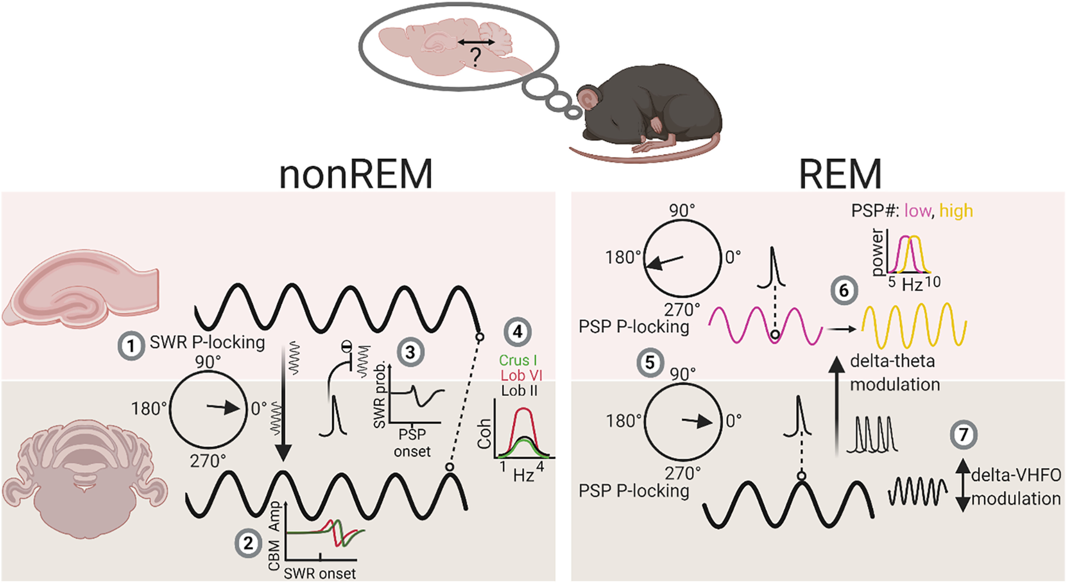
Figure 8.

Summary diagram of main findings illustrating sleep stage-specific physiological events and interactions within the cerebello-hippocampal network. During non-REM, hippocampal SWRs are phase-locked to the cerebellar δ oscillation (1) and drive modulation of cerebellar LFP activity, at shortest latency in Lob VI (2). In addition, PSPs recorded in the cerebellum are associated with reduced SWR probability of occurrence. Finally, overall coherence within the δ frequency range is highest between hippocampus and Lob VI compared with other cerebellar lobules. During REM, PSPs are significantly phase-locked to both the trough of hippocampal theta (purple line) and the peak of cerebellar δ oscillations (black line) (5). Cerebellar δ oscillations and associated PSPs modulate the frequency of hippocampal theta rhythms (from ∼7.5 Hz [low, purple line] to 8 Hz [high, yellow line]) (6). Additionally, within the cerebellum, δ oscillations can modulate activity within the VHFO range during REM (7). P-locking, phase-locking. The multisite LFP recording technique used in our study allowed us to survey a large spatial extent of the cerebellum (hemispheric, Crus I; dorsal vermis, Lob VI; and ventral vermis, Lob II/III) alongside hippocampal LFP, which would be difficult to achieve using alternative methods. However, this methodology precluded layer-specific or local spiking activity measurement. Future studies directly measuring spiking activity across both regions during sleep will be essential to fully understand the functional significance of the dynamics observed at the LFP level. Created with www.BioRender.com.
