. 2022 Feb 10;11:e71928. doi: 10.7554/eLife.71928

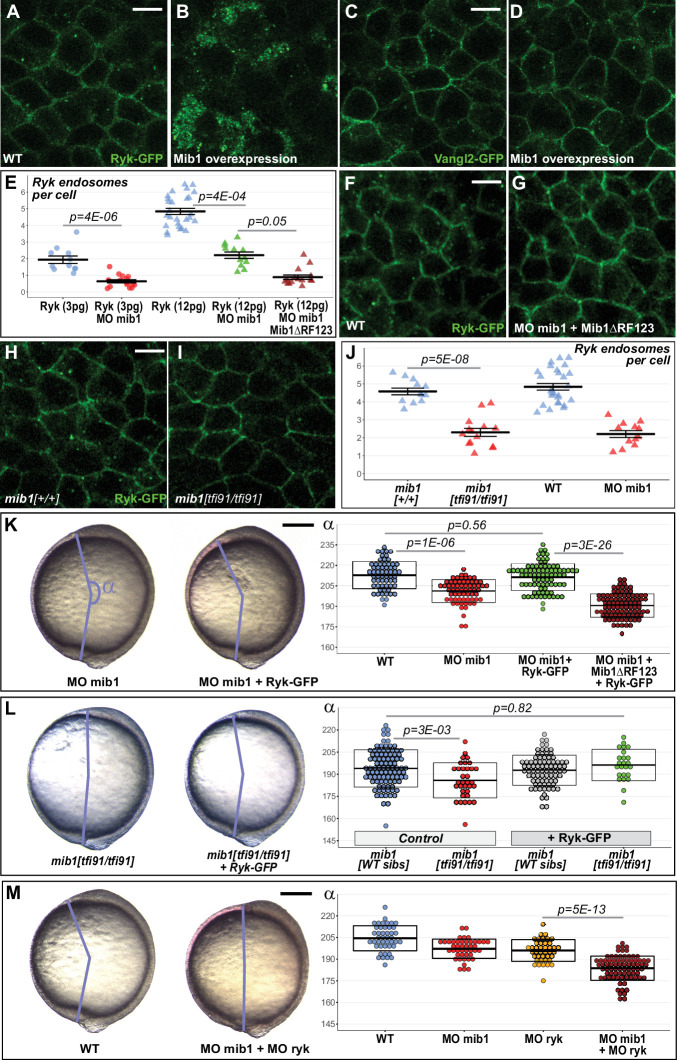
Figure 3. Mib1-mediated Ryk endocytosis controls Convergent Extension movements.

(A–D) WT mib1 RNA injection triggers Ryk internalization in 20/21 embryos (B) but has no effect on Vangl2 localization (D, n = 23). (E–G) Mib1 morpholino injection reduces the number of Ryk endosomes that are present upon injection of Ryk-GFP RNA. Increasing the dose of Ryk-GFP RNA restores endosome number in mib1 morphants but not in embryos coinjected with Mib1ΔRF123. (H–J) The number of Ryk endosomes that are present upon injection of Ryk-GFP RNA (12 pg) is reduced in mib1 null mutants. mib1 morphant data from panel E are shown again for comparison. (K) Ryk-GFP RNA (12 pg) rescues axis extension in mib1 morphants but not in embryos coinjected with Mib1ΔRF123. (L) Similarly Ryk-GFP injection rescues axis extension in mib1tf91 mutants. (M) Ryk morpholino injection aggravates mib1 morphant axis extension phenotypes. (A–D,F,G,H,I) dorsal views of 90% epiboly stage embryos, anterior up, scalebars 10 µm. (K,L,M) Lateral views of bud stage embryos, anterior up, scalebars 200 µm. In (E,J) each data point represents the mean number of endosomes for 20 cells from a single embryo. For comparison J again includes the mib1 morphant from panel E. Bars represent mean values ± SEM. In (K,L,M) boxes represent mean values ± SD. See Figure 3—source data 1 for complete statistical information.

Figure 3—source data 1. Complete statistical information for the experiments reported in Figure 3 and Figure 3—figure supplement 5.

Figure 3.

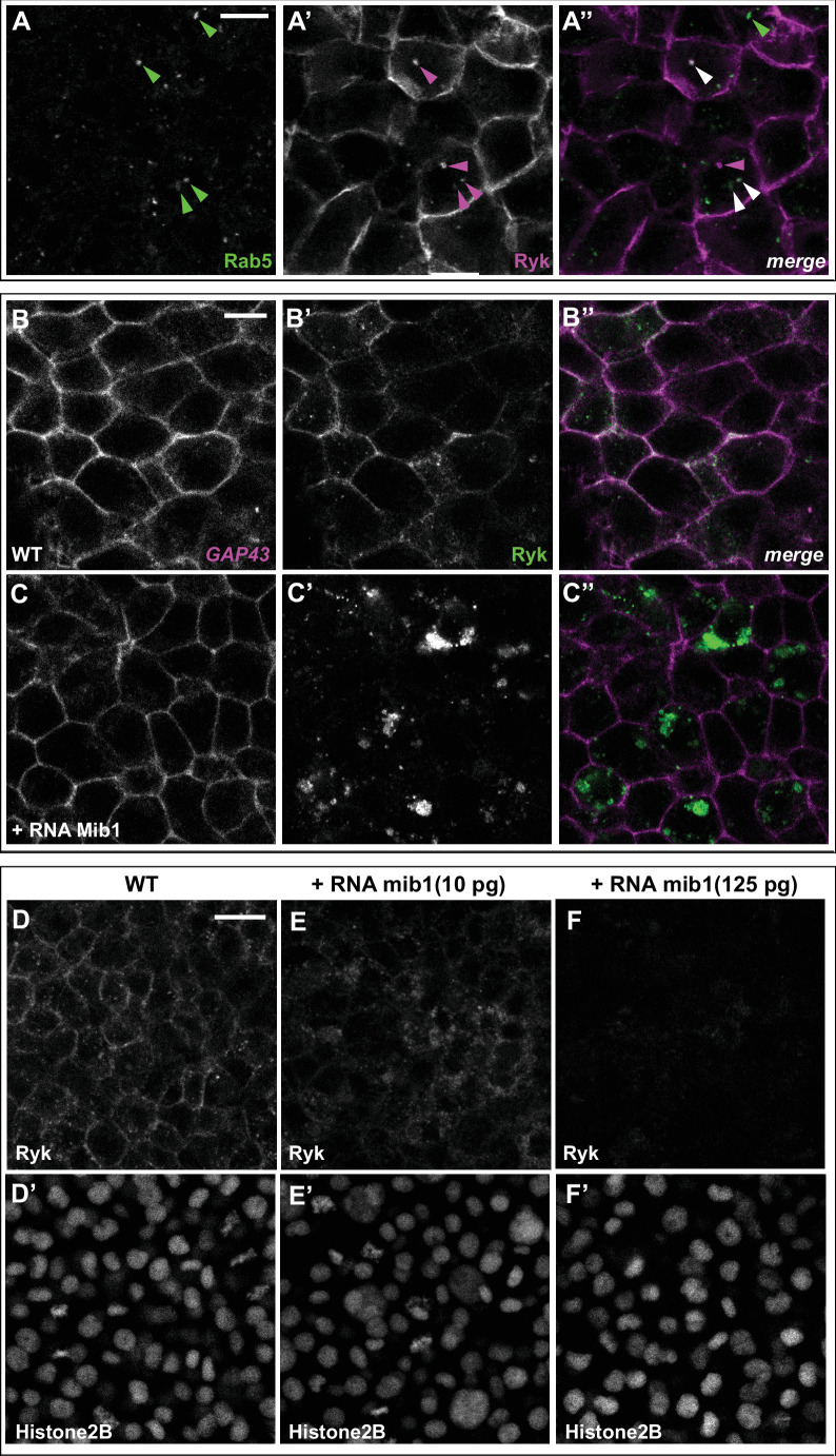
Figure 3—figure supplement 1. Mib1 promotes Ryk internalization and degradation.

Figure 3—figure supplement 1.

(A) Coinjection of RNAs encoding Rab5-GFP and Flag-Ryk-Myc reveals that 70.7% of Ryk-expressing intracellular compartments are positive for the early endosomal marker Rab5 (n = 75 cells from eight embryos analyzed). Arrowheads in A’’ indicate compartments that are positive for Rab5 only (green), Ryk only (magenta), or present both markers (white). (B,C) Mib1 overexpression promotes the internalization of Ryk-GFP but not the one of the plasma membrane marker GAP43-RFP (n = 4). (D,E) RNAs encoding Ryk-GFP and Histone2B-mRFP were injected with increasing amounts of mib1 RNA. While a low dose of Mib1 relocalizes Ryk from the plasma membrane to intracellular compartments (n = 6), high amounts of Mib1 cause an overall loss of Ryk signal (n = 6). D’-F’, The Histone2B-mRFP signal was used to ascertain that embryos had received a comparable amount of injected material. (D-F and D’-F’) are sum projections of three consecutive slices from confocal stacks. All pictures depict dorsal views of 90% epiboly stage embryos, anterior up. Scalebars: 10 µm in A-C, 20 µm in D-F.
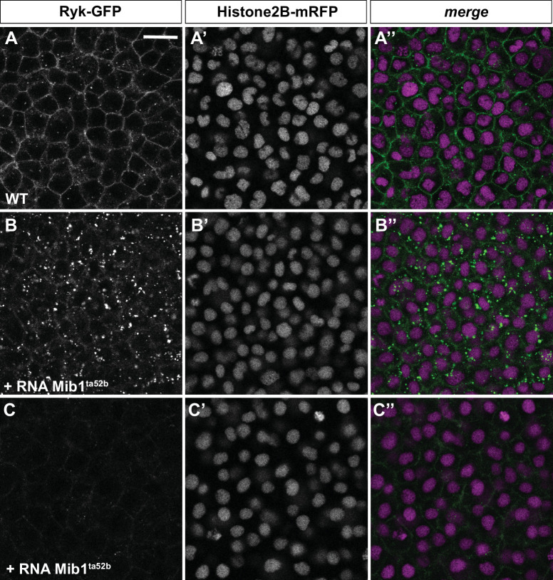
Figure 3—figure supplement 2. Mib1ta52b overexpression promotes Ryk internalization.

Figure 3—figure supplement 2.

(A–C) Microinjection of RNA encoding the Mib1ta52b mutant protein induces Ryk internalisation (B, n = 16/23 embryos) or degradation (C, n = 7/23 embryos) as compared to WT controls (A, n = 21). Dorsal views of 90% epiboly stage embryos, anterior up. The Histone2B-mRFP signal (A’-C’) was used to ascertain that control and Mib1ta52b-expressing embryos had received comparable amounts of injected material. Scalebar: 20 µm.
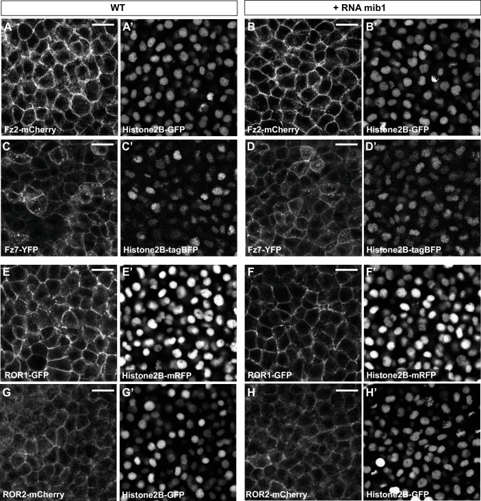
Figure 3—figure supplement 3. Mib1 overexpression does not affect Frizzled/Ror localization.

Figure 3—figure supplement 3.

(A–D) Mib1 overexpression has no effect on the localization of the Wnt receptors Frizzled2 (Fz2, n = 6) or Frizzled7 (Fz7, n = 8). (E–H) Mib1 overexpression has no effect on the localization of the Wnt-binding receptor tyrosine kinases ROR1 (n = 7) or ROR2 (n = 10). All pictures depict dorsal views of 90% epiboly stage embryos, anterior up. (G,H) are sum projections of three consecutive confocal slices. A’-H’ Display the signal for fluorescently tagged Histone2B constructs that were coinjected to ascertain that control and mib1-expressing embryos had received a comparable amounts of injected material. Scalebars: 20 µm.
Figure 3—figure supplement 4. Mib1 loss of function impairs Ryk endocytosis.

Figure 3—figure supplement 4.

(A,B) mib1 morpholino (MO mib1) injection reduces Ryk endocytosis. (C,D) A more pronounced inhibition of Ryk endocytosis is observed upon coinjection of MO mib1 and RNA encoding dominant-negative Mib1 (mib1DRF123). (E,F) Ryk endocytosis is reduced in mib1tfi91 mutant embryos. All pictures depict dorsal views of 90% epiboly stage embryos, anterior up. A’-F’, The Histone2B-mRFP signal was used to ascertain that control and mib1-depleted embryos had received a comparable amount of injected material. Embryos depicted in A-F were injected with 12 pg Ryk-GFP RNA. (C-F) correspond to the display items also shown in Figure 3F–I. Scalebars: 10 µm.
Figure 3—figure supplement 5. mib1 morphant defects are not rescued upon vangl2 overexpression.
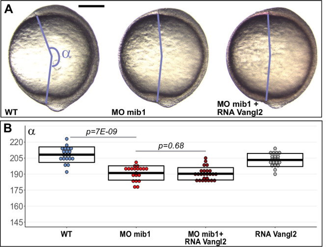
Figure 3—figure supplement 5.

(A,B) vangl2 RNA injection does not rescue axis extension in mib1 morphants. (A) Lateral views of bud stage embryos, anterior up, dorsal to the right. Scalebar 200 µm. In (B) boxes represent mean values ± SD. See Figure 3—source data 1 for complete statistical information.