Figure 2.

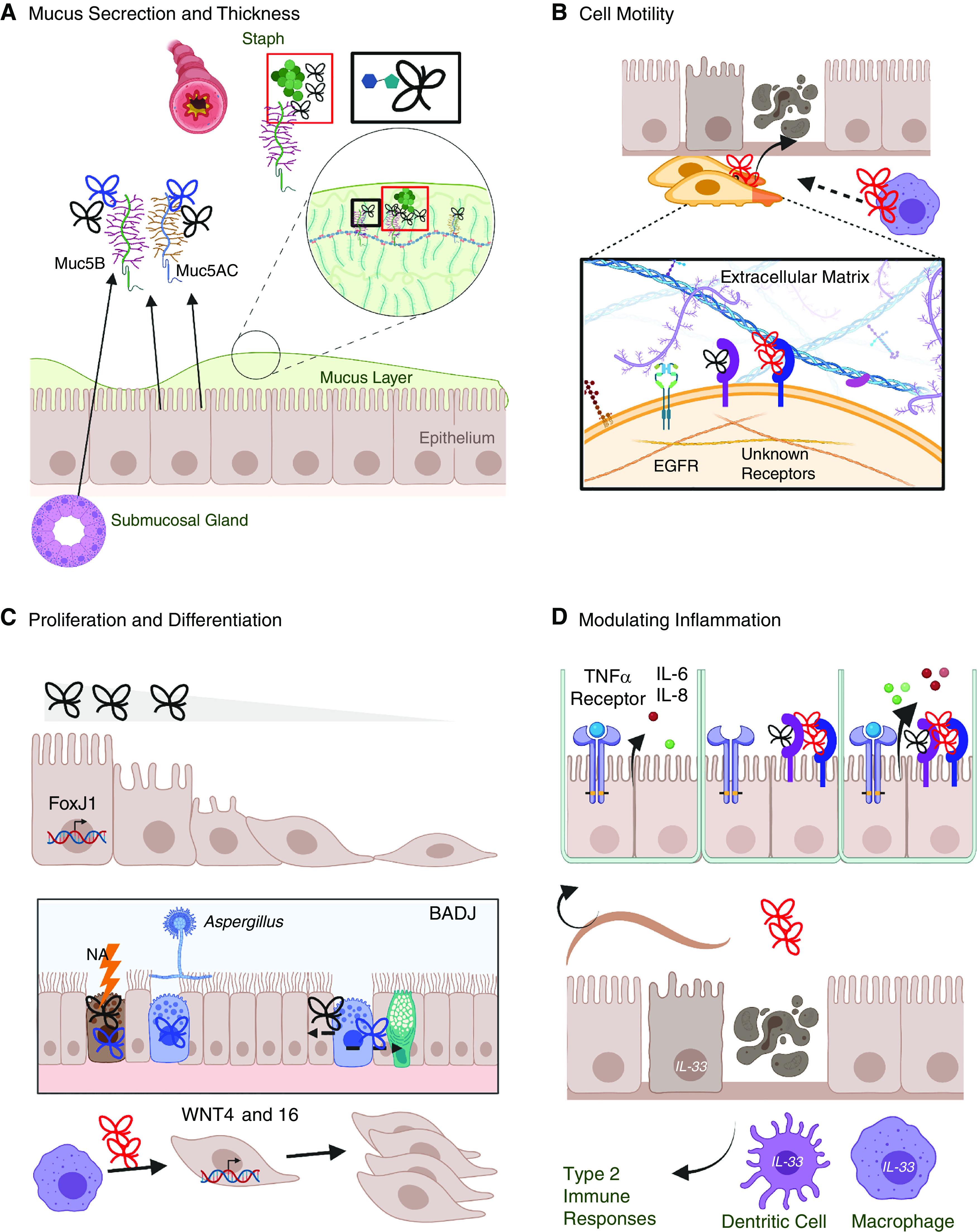
Contribution of TFFs to mucosal barrier function and repair. (A) TFFs bind the terminal disaccharide of mucin glycoproteins to thicken the mucous layer (box outlined in black), which can constrict the airway. TFF1 (blue) and TFF3 (black) are associated with Muc5AC (epithelial) and Muc5B (epithelial and submucosal). TFF1 has been found around Staph, and Muc5B is required for bacterial clearance in the lung (box outlined in red). (B) Cell migration into a damaged site is required for tissue repair. TFF2 (red) and TFF3 (black) bind with unknown receptors to enhance cell motility synergistically with EGF (epidermal growth factor) and extracellular matrix components. (C) TFF3 and FoxJ1 transcription induce ciliogenesis. In the BADJ, NA injury or allergen exposure induces TFF1 and/or TFF3 in club cells, which can differentiate into epithelial or goblet cells. TFF2 from macrophages induces WNT epithelial expression, which is associated with cell proliferation. (D) TFFs contribute to immune function, including enhancing TNFα-evoked release of IL-6 and IL-8; TFF2-dependent induction of IL-33 in epithelial cells, dendritic cells, and macrophages; type 2 responses; and worm clearance during helminth infection. BADJ = bronchoalveolar duct junctions; EGFR = EGF receptor; NA = naphthalene; Staph = Staphylococcus aureus.
