
Figure 1.
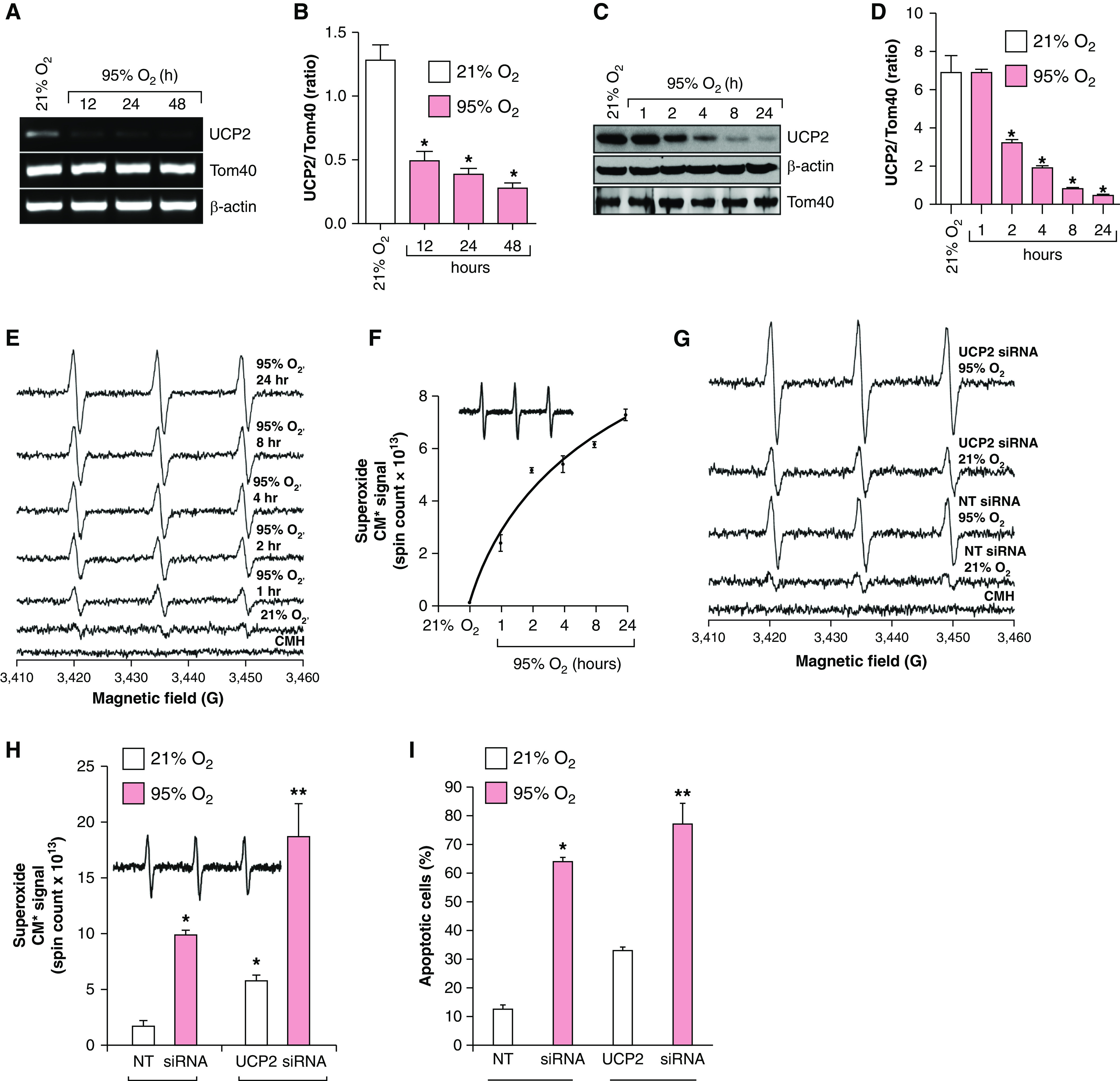
Hyperoxia decreases UCP2 (uncoupling protein 2) expression, and loss of UCP2 increases superoxide anion (O2•−) generation and apoptosis in lung cells. (A) MLE-12 cells (n = 3) were exposed to 21% or 95% O2 for the indicated time, and total RNA was isolated and analyzed for UCP2 and Tom40 (mitochondrial protein control) expression and normalized for β-actin. (B) Densitometry of A. *P < 0.05, hyperoxia versus normoxia (ANOVA). (C) Extracts from MLE-12 cells (n = 3) were exposed to normoxia or hyperoxia and analyzed for UCP2 expression through Western blotting using its specific antibody. (D) Densitometry of C. *P < 0.05, hyperoxia versus normoxia (ANOVA). (E) MLE-12 cells (n = 3) were exposed to 21% O2 or 95% O2 for the indicated periods, and O2•− generation was determined through electron paramagnetic resonance (EPR) by using 1-hydroxy-3-methoxycarbonyl-2,2,5,5-tetramethylpyrrolidine hydrochloride (CMH) as described in the Methods. (F) Superoxide production (CM*, nitroxide radical) over time in hyperoxia (spin counts over time). (G) MLE-12 cells (n = 3) were transfected with 100-nM nontargeted (NT) siRNA or UCP2 siRNA and exposed to 21% O2 or 95% O2 for 24 hours. The O2•− level was determined by using EPR. (H) Total spin counts from G are plotted as a bar graph. *P < 0.01, 95% O2 + NT siRNA versus 21% O2 + NT siRNA; and **P < 0.01, 95% O2 + UCP2 siRNA versus 21% O2 + UCP2 siRNA. (I) MLE-12 cells (n = 3) were transfected with 100-nM NT siRNA or UCP2 siRNA and exposed to 21% or 95% O2 for 24 hours, and apoptosis was determined by using an annexin V binding assay. *P < 0.05, 21% O2 + NT siRNA versus 95% O2 + NT siRNA; and **P < 0.05, 21% O2 + UCP2 siRNA versus 95% O2 + UCP2 siRNA.