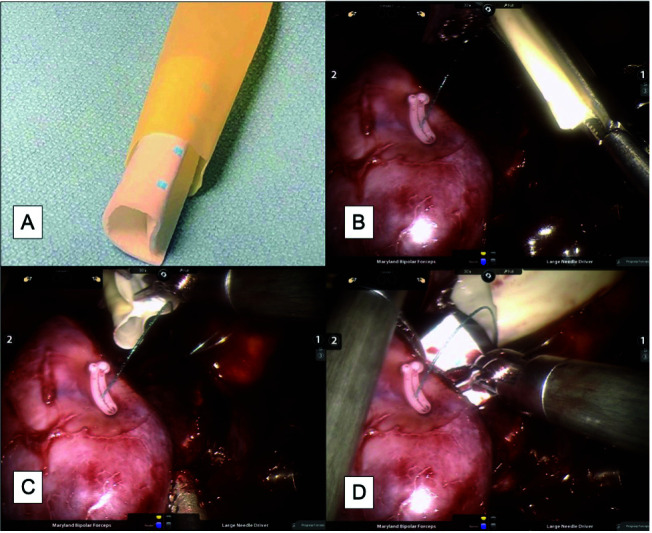
Fig. 1.

(A) Preparation and deployment of a PEG-coated patch into a renal mass defect. The patch is rolled with the adhesive side facing inward and placed into the cut 5th finger of a sterile glove. (B) The assistant deploys the glove finger into the field using a laparoscopic grasper. (C, D) The surgeon can manipulate the hemopatch onto the defect without contact with surrounding tisues and fluids.
