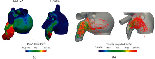
Figure 3.

(a) Three-dimensional maps of endothelial cell activation potential (ECAP) for a transient ischemic attack or cerebrovascular accident patient (left) and a control case (right). High ECAP values (red areas) indicate a higher risk of thrombus formation due to low velocities and complex blood flow. (b) These velocities as well as the complexity of the flow within the LAA can be visualized with the streamlines for a thrombus case (left), while in a control case (right) flow remains in the ostium and does not reach the tip part of the LAA.
