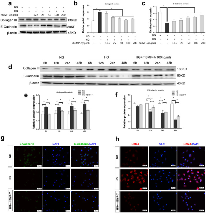
Fig. 3. BMP-7 inhibited high glucose-mediated Collagen-III expression and preserved cell phenotype of NRK-52E cell.
Representative a Western blot and quantitative data were revealed the expression of b Collagen-III, c E-cadherin, in NRK-52E cells under normal-glucose condition, high glucose condition with either vehicle or recombinant human BMP-7(rhBMP-7) at different dosages,*P < 0.05 (n = 3). Representative d Western blot and quantitative data were revealed the expression of e Collagen-III, f E-cadherin, in NRK-52E cells under normal-glucose condition, high glucose condition with rhBMP-7(100 ng/ml) at different time, *P < 0.05 (n = 3). g Representative micrographs show immunofluorescence staining of E-cadherin in different groups. DAPI staining(blue) represents cell nucleus, Bar = 50 um. h Representative micrographs show immunofluorescence staining of α-SMA in different groups. DAPI staining(blue) represents cell nucleus, Bar = 50 um.

