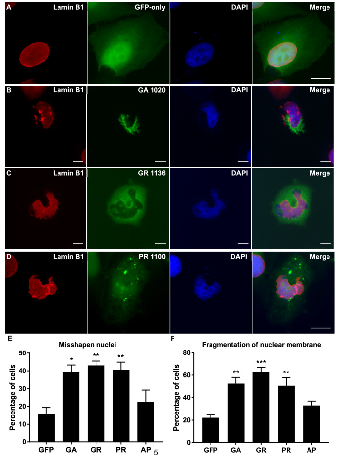
Figure 1.
Abnormal nuclei in DPR-expressing cells. (A–D) Lamin B1 was used as a marker for the nuclear membrane (red). HeLa cells transfected with GFP-tagged DPR constructs or empty pEGFP-N1 vector as a GFP-only control were fixed and stained 48 h post-transfection. Nuclei were frequently “horseshoe-shaped” or excessively folded in cells containing GA1020 (P = 0.0131), GR1136 (P = 0.0052) or PR1100 inclusions (P = 0.0096). AP1024 did not cause nuclear abnormalities (data not shown; P = 0.6797). In addition, the nuclear membrane is fragmented in cells expressing GA1020 (P = 0.0047), GR1136 (P = 0.0006) or PR1100 (P = 0.0071), but not AP1024 (data not shown; P = 0. 3865). DAPI was used to stain nuclei (blue). Scale bars indicate 15 μm. (E, F) Graphs shows percentage of transfected cells with misshapen nuclei (E) or fragmented nuclear membranes (F). n = 3, with a minimum of 30 cells analysed for each independent replicate. Data was analysed by one-way ANOVA with Dunnett’s multiple comparison test. Error bars indicate SEM.

