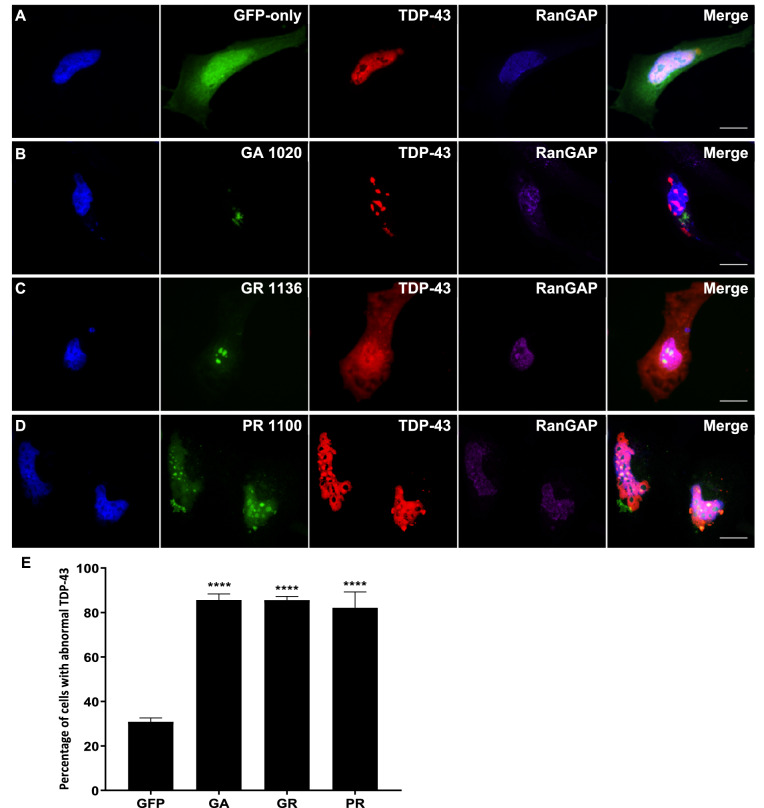
Figure 6.
DPR-induced mislocalisation of TDP-43 and RanGAP occur together. (A–D) HeLa cells co-transfected with mCherry-tagged human TDP-43 (red) and GFP-tagged DPR constructs or empty pEGFP-N1 vector as a GFP-only control were fixed and stained 48 h post-transfection. RanGAP was stained using a Cy5-conjugated antibody (purple). Cells exhibiting DPR-induced TDP-43 mislocalisation typically also exhibited granular accumulation of RanGAP in the nucleus. Scale bars indicate 15 μm. (E) The mean percentage of cells with abnormal TDP-43 that also exhibited granular accumulation of RanGAP in the nucleus was significantly increased by GA1020 (P < 0.0001), GR1136 (P < 0.0001), and PR1100 (P < 0.0001). n = 3, with a minimum of 30 cells analysed for each independent replicate. Data was analysed by one-way ANOVA with Dunnett’s multiple comparison test. Error bars indicate SEM.

