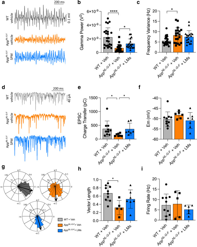
Fig. 2. LMs partially restore gamma oscillation power and synchronization in AppNL-G-F mice.
a Representative traces of gamma oscillations in hippocampal brain slices are shown in wild-type (WT) and AppNL-G-F mice given vehicle (Veh) and AppNL-G-F mice treated with lipid mediators (LMs). b Bar graphs of the gamma oscillation power in the three animal groups. Treatment with LMs partially restored the reduced gamma power observed in AppNL-G-F mice. c Bar graphs of gamma oscillations described in b. There was an increase in frequency variance in the App NL-G-F mice, but the LM treatment did not reduce this. d Representative traces of excitatory postsynaptic currents (EPSCs) from fast-spiking interneurons (FSNs) in each condition. e Bar graphs showing EPSC charge transfer in FSNs from each condition. Treatment with LMs restored the reduction in EPSC charge transfer observed in AppNL-G-F mice. f Bar graphs of FSN membrane potential (Em) from each condition. There was no difference in the Em between the three groups. g Polar-plots showing the distribution of AP phase-angles from the experimental conditions described in A. h Bar graphs of resulting vector length derived from concomitant recordings of gamma oscillations and FSN (AP) distribution for each experimental condition. There was a decrease in the vector length in the AppNL-G-F mice, but the LM treatment did not restore this. i Bar graphs of FSN firing rate obtained from H. Kruskal-Wallis and one-way ANOVA tests were used for group comparisons. Data are presented as mean ± S.E.M. *P < 0.05, **P < 0.01, ***P < 0.001.

