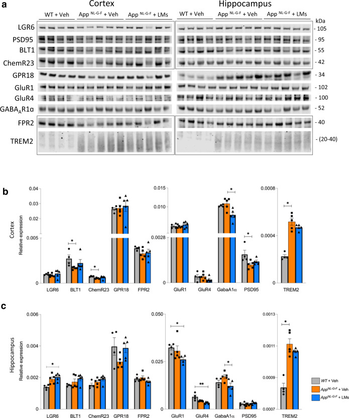
Fig. 4. Receptors and synaptic markers are altered in AppNL-G-F mice.
(a–c) Analysis of receptors for pro-resolving lipid mediators (LMs) (LGR6, BLT1, ChemR23, GPR18 and FPR2), glutamate and GABA receptors (GluR1, GluR4, GABAA1α), a synaptic marker (PSD95), and an inflammation marker (TREM2), was performed by Western blot in cortex and hippocampus of WT + Veh, AppNL-G-F + Veh and AppNL-G-F + LMs mice. Representative blots (a) and quantitative analysis (b, c) are shown (markers from the same blots are indicted by rectangles, and complete Western blots are shown in Supplementary Fig. 2). Data are presented as mean ± SEM and analysed by Kruskal-Wallis one-way analysis of variance test with Dunn’s post hoc test (*P < 0.05, **P < 0.01, ***P < 0.001, ****P < 0.001) between treatment groups for each marker (n = 4 - 5 mice/group). Data are presented as mean ± S.E.M. LGR6 leucine-rich repeat containing G-protein coupled receptor 6, BLT1 leukotriene B4 receptor, ChemR23 chemokine-like receptor 1, GPR18 G-protein-coupled receptor 18, FPR2 formyl peptide receptor 2, TREM2 triggering receptor expressed on myeloid cells 2, GluR1 glutamate receptor 1, GluR4 glutamate receptor 4, GABAA1α gamma-aminobutyric acid A receptor subunit 1α, PSD95 postsynaptic density protein 95, FC fear conditioning, NOR novel object recognition, DI discrimination index.

