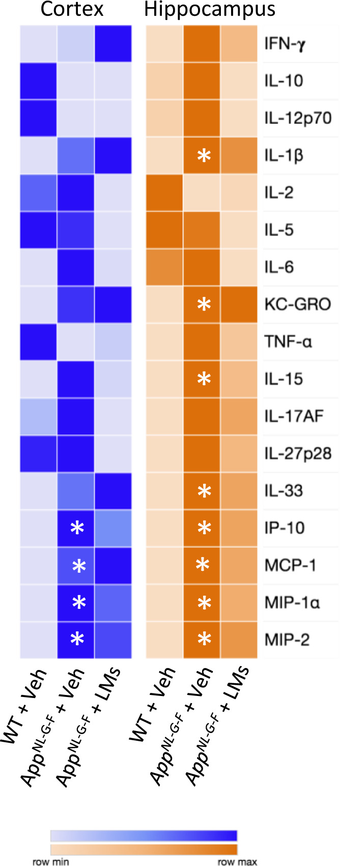
Fig. 5. Cytokines and chemokines are altered in the brain of AppNL-G-F mice.

Cytokines and chemokines were analysed in homogenates of cerebral cortex and hippocampus by multi-immunoassay (Meso Scale v-plex). Rows in the heatmap represent cytokines and chemokines and columns represent treatment groups. The colours represent median of normalized concentration values. n = 5 mice/group. Asterisks denote statistical significance between wild-type (WT) and AppNL-G-F mice given vehicle (Veh) using Kruskal-Wallis with Dunn’s post hoc test. IFN-γ interferon-γ, IL interleukin, IP-10 interferon-γ-induced protein 10, KC-GRO keratinocyte chemoattractant/human growth-regulated oncogene, MCP-1 monocyte chemoattractant protein, MIP macrophage inflammatory protein, TNF-α tumour necrosis factor-α.
