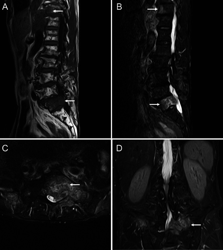
Fig. 2.
Magnetic resonance imaging on the first admission suggests bone metastasis with infiltration in the spinal canal. A T1-weighted image shows low signal intensity in the thoracic and sacral vertebra (arrow). B T2-weighted short TI inversion recovery (T2W-STIR) image shows high signal intensity in thoracic and sacral vertebra (arrow), suggesting bone metastasis. C, D T2W-STIR image (C axial), (D coronal) showing tumor infiltration in the spinal canal

