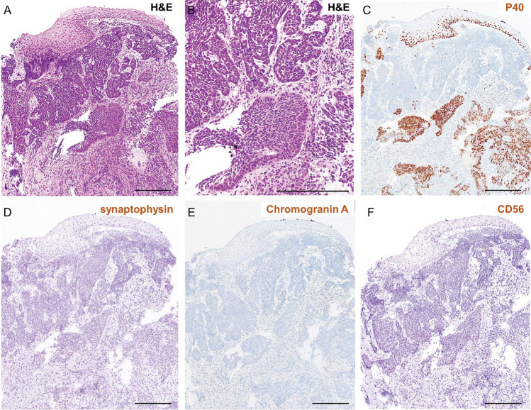
Fig. 4.
Poorly differentiated squamous cell carcinoma diagnosed by pathological analysis. A, B Hematoxylin and eosin (H&E) staining in low (A) and high (B) magnification. Atypical cells with a high nuclear–cytoplasmic (N/C) ratio proliferate under the epithelium in an irregular honeycomb pattern. Scale bar: 100 μm. C P40 immunostaining revealed many atypical P40 positive cells and some P40 negative cells. D–F Neuroendocrine marker immunostaining is shown. D Synaptophysin and E Chromogranin A and F CD56 immunostaining revealed that almost all the atypical cells were negative for these markers

