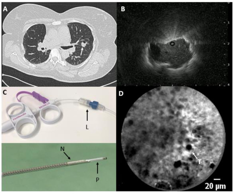
Figure 2.
(A) Chest CT scan with a lung tumour in the left upper lobe (arrow) and (B) radial endobronchial ultrasound image with an eccentric tumour visualisation. (C) Preloading of the needle:after adjusting the luer lock (L) on the 18G Broncus needle, the confocal miniprobe is advanced through the luer lock, positioning the tip of the probe (P) 4 mm past the needle tip (N). (D) In-vivo needle-based confocal laser endomicroscopy image at the tip of the needle showing real-time pleomorphic enlarged tumour cells (T) representing a sarcoma metastasis.

