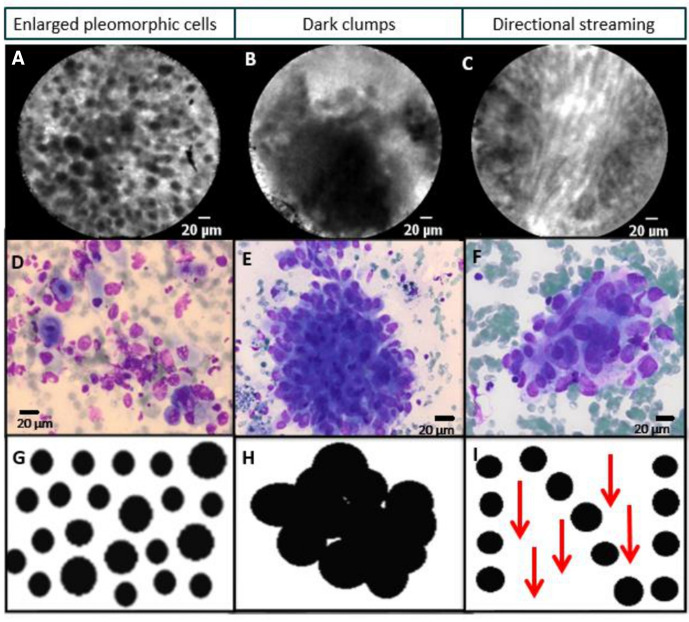
Figure 3.
(A–C) Real-time needle-based confocal laser endomicroscopy (nCLE) imaging of different lung tumours demonstrating the two ‘static’ nCLE malignancy criteria (enlarged pleomorphic cells and dark clumps) and the ’dynamic’ phenomenon of directional streaming (online example). (D–F) Corresponding cytology of the fine needle aspirate representing squamous cell carcinoma, adenocarcinoma and sarcoma metastasis. (G–I) Schematic display of malignant nCLE features.

