Figure 4.
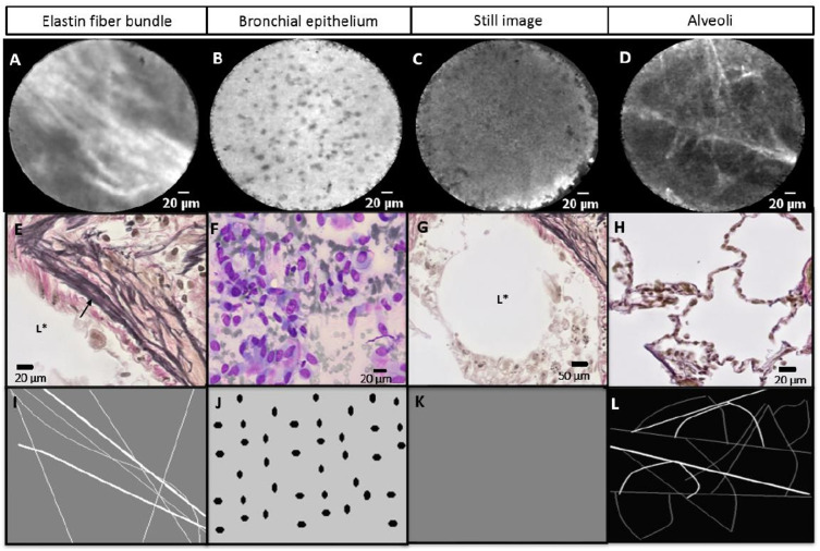
(A–D) Real-time needle-based confocal laser endomicroscopy (nCLE) imaging at the needle tip showing three nCLE airway criteria (elastin fibres, bronchial epithelium and still image) and the alveoli of the lung parenchyma. (E–H) Histology (E, G and H) and cytology (F) of the different structures in the airway and lung parenchyma. (I–L) Schematic display of the airway (I–K) and lung parenchyma (L) nCLE features. (A) Autofluorescent elastin fibre bundles (indicated in E by arrow) along the lumen of the airway (indicated in E by L*). (B) Small homogeneous and equally distributed cells representing the bronchial epithelium (F). (C) Still nCLE image as the result of the CLE-probe being advanced in the lumen of a larger airway (indicated in G by L*) without touching the airway wall. (D) Autofluorescent alveoli with a hexagonal architecture (H). Scale bar: (A–F and H) 20 µm and (G) 50 µm.

