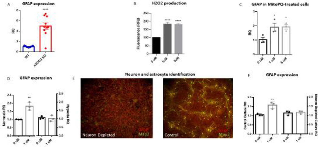
Figure 4.

Neuronal SOD2 deficiency in vivo and neuronal mitochondrial oxidative stress in vitro induces astrogliosis. (A) GFAP transcription was found to be increased in the cortex of nSOD2 KO mice at 2 months of age as compared to WT littermates in a whole genome array and this finding was verified via RT-PCR (unpaired t test, n=4-5, p<0.0001). (B) The selective mitochondria-target redox cycler MitoPQ increases ROS production in rat primary cortical cells at 1 and 5 μM within 24 hours as measured by amplex red (one-way ANOVA with Dunnett’s multiple comparison, p<0.0001). (C) Following a one week incubation, 1 and 5 uM MitoPQ induces astroglyosis as measured by GFAP gene expression (three independent trials, one-way ANOVA with Dunnett’s multiple comparison, p=0.0463 (1uM) and p=0.0413 (5uM)). (D) This induction of GFAP is abated when cells are grown under hypoxic conditions (unpaired t test, n=3 independent trials, p=0.0008 (normoxia) p=0.5213 (hypoxia). (E) ICC of GFAP (red) and Map2 (green) confirms the establishment of neuronally-depleted cortical cultures as compared to side-by-side controls. (F) Neuronal depletion prevents MitoPQ-induced astrogliosis as evidenced by GFAP gene expression (unpaired t test, n=3 independent trials, p=0.028 (control), p=0.9186 (neuron depleted)
