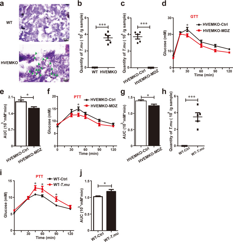
FIG 1.
The murine protozoan T. musculis promotes gluconeogenesis. (a) The cecal contents of WT and HVEM KO mice were fixed, stained with hematoxylin and eosin, and visualized microscopically. Green arrowheads indicate protozoa. Scale bar, 10 μm; n = 3. (b) Total number of T. musculis protozoa in the cecal contents of the indicated mice. n = 5. (c to g) Eight-week-old male HVEM KO mice were treated with vehicle or 2.5 g/liter metronidazole (MDZ) in drinking water for 1 week. The number of T. musculis presented in the cecal content was enumerated before (ctrl) and after MDZ treatment (c). Glucose tolerance testing (GTT) (d and e) or pyruvate tolerance testing (PTT) (f and g) were performed. AUC, the area under the curve. n = 5 mice per group. (h to j) T. musculis protozoa isolated from HVEM KO mice were transferred to T. musculis-free WT mice (1 × 10⁶/mouse). The number of T. musculis colonized in the WT recipient mice was calculated (h), and PTT was performed (i and j). n = 5 mice per group. The experiments were repeated at least two times. The data represent means ± the SEM. *, P < 0.05; **, P < 0.01. T.mu, T. musculis.

