Figure 1.
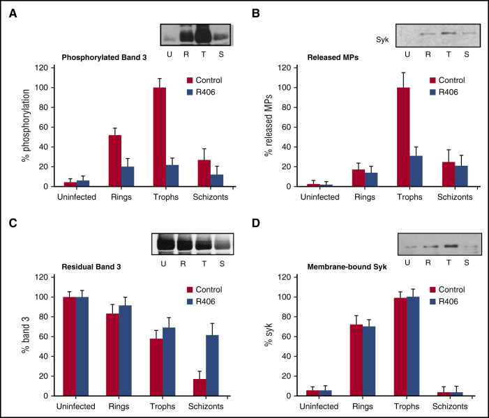
Abrogation of the P falciparum–induced erythrocyte membrane modifications by SYK inhibitors at different P falciparum lifecycle stages. Synchronized P falciparum cultures were supplemented with SYK inhibitor R406 (1.0 µM) at ring stage (24 hours after invasion). (A) Band 3 tyrosine phosphorylation levels are expressed as percentages of band 3 maximal phosphorylation measured at trophozoite stage and were normalized by the content of band 3 at each stage (% phosphorylation). The insert shows a representative western blot of tyrosine phosphorylated band 3 in the absence of inhibitors in uninfected RBCs (U); ring-infected RBCs (R); trophozoite-infected RBCs (T); and schizont-infected RBCs (S). (B) Number of MPs released from control and R406-treated cells. Values are expressed as a percentage of the maximal number of released MPs measured at trophozoite stage (% Released MPs). The insert shows a representative anti-Syk western blot of MPs released at different stages (U, R, T, S). (C) Band 3 content measured in control and R406-treated cells. Values are expressed as percentage of the band 3 measured at different stages using uninfected cells as reference (% band 3). The insert shows a representative western blot of band 3 at different stages (U, R, T, S). (D) Membrane-bound Syk measured in control and R406-treated cells. Values are expressed as a percentage of the maximal levels of bound Syk measured at trophozoite stage (% Syk). The insert shows a representative western blot of Syk at different stages (U, R, T, S). Data are the average of 6 independent experiments ± standard deviation (SD). Nitrocellulose membranes were stained with rabbit anti-Syk (Cell Signaling Technology, Inc., CA) diluted to 1:1000, and with goat anti-rabbit IRDye 800CW (LI-COR), diluted to 1:50 000. Quantitative densitometry analysis was performed using Odyssey V3.0 software. Photomicrographs were acquired using Odyssey from LI-COR, setting 300dpi as definition.

