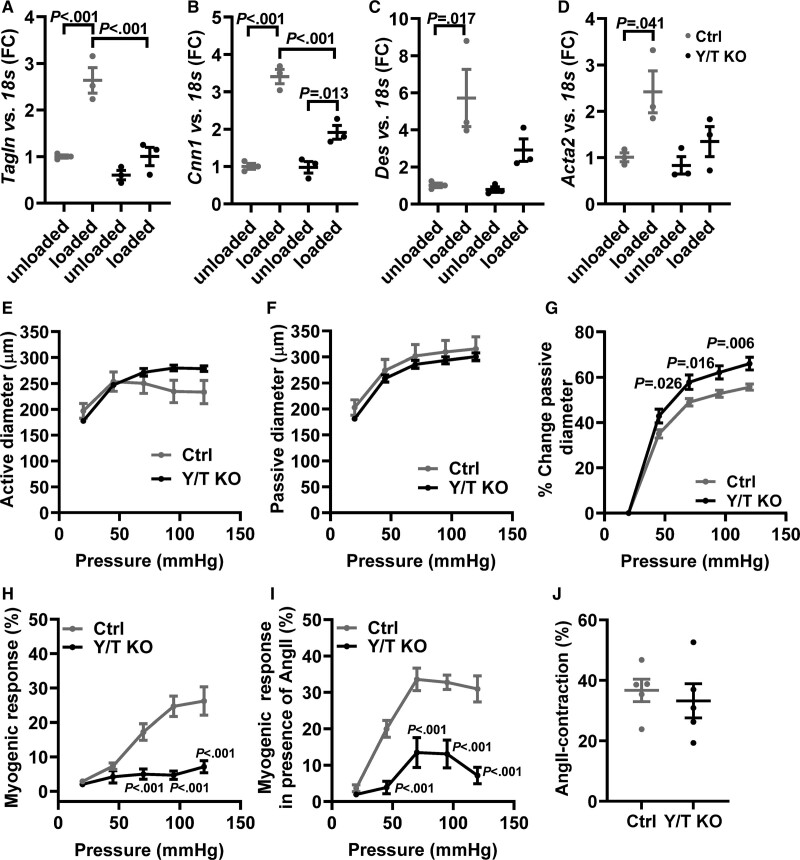
Figure 3.
Reduced expression of stretch-induced contractile markers and decreased myogenic response in Y/T KO (YAP/TAZ [yes-associated protein 1/WW domain containing transcription regulator 1] knockout) mice. The portal veins (n=3 in all groups and targets) were kept in organ culture for 24 h either stretched with gold weight (loaded) or not stretched (unloaded). Reverse transcription-quantitative polymerase chain reaction analysis of selected smooth muscle markers in portal veins from Y/T KO and Ctrl (control) mice: (A) Tagln, (B) Cnn1, (C) Des, and (D) Acta2. E–J, Mesenteric arteries were mounted in a pressure myograph. E, Intraluminal pressure was increased systematically, and active vessel diameter was recorded in Ca2+ containing HEPES-buffered Krebs solution. F, Passive vessel diameter was measured in Ca2+-free solutions. G, The relative change in passive diameter was used as an indicator of vascular distensibility. H, Myogenic response was calculated as the relative difference between active and passive diameter. I, A single dose of 100 nmol/L Ang II (angiotensin II) was added to the preparations followed by gradual increase in intraluminal pressure. Active diameter was monitored, and myogenic response was calculated (E–I; Ctrl, n=6; Y/T KO, n=6). J, Peak transient contraction relative to baseline after 100 nmol/L Ang II stimulation (Ctrl, n=5; Y/T KO, n=5). All data are presented as mean±SEM. FC indicates fold change.

