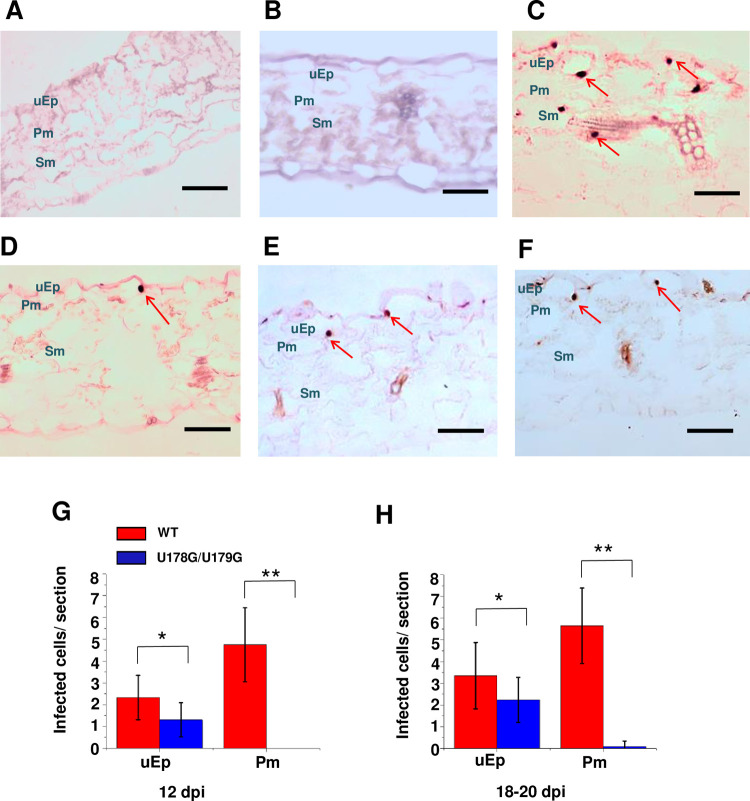
Fig 7. U178G/U179G fails to exit epidermal cells in rub-inoculated leaves.
Transverse section (12 μm) in situ hybridization of (A) mock inoculated leaves (negative control), (B) leaves inoculated with replication defective A271G/C273G (negative control), (C) wild type PSTVd (positive control), and (D-F) U178G/U179G. Images for PSTVd WT and U178G/U179G are representative of more than 200 sections. Purple dots (red arrows) are viroid hybridization signals in nuclei. uEp, upper epidermis; Pm, palisade mesophyll; Sm, spongy mesophyll; lEp, lower epidermis. Bars = 100 μm. (G and H) Number of infected cells per leaf section (-1 x 0.15 mm) in the upper epidermis (uEp) or adjacent palisade mesophyll (Pm) of plants inoculated with WT PSTVd (red) or U178G/U179G (blue) at 12 dpi (G) and 18–20 dpi (H). Data were compiled from 40 sections obtained from 20 infected plants. Asterisks indicate significant differences (p < 0.05*; p < 0.01**) as determined by Student’s t test. Bars indicate standard error of the mean.

